Review
. 2015:84:291-323.
doi: 10.1146/annurev-biochem-060614-034316. Epub 2015 Mar 12.

Mechanisms and Regulation of Alternative Pre-mRNA Splicing

Affiliations
Review

Mechanisms and Regulation of Alternative Pre-mRNA Splicing

Yeon Lee et al. Annu Rev Biochem. 2015.

Abstract

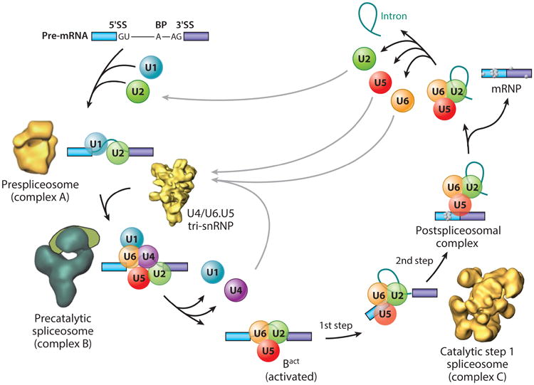
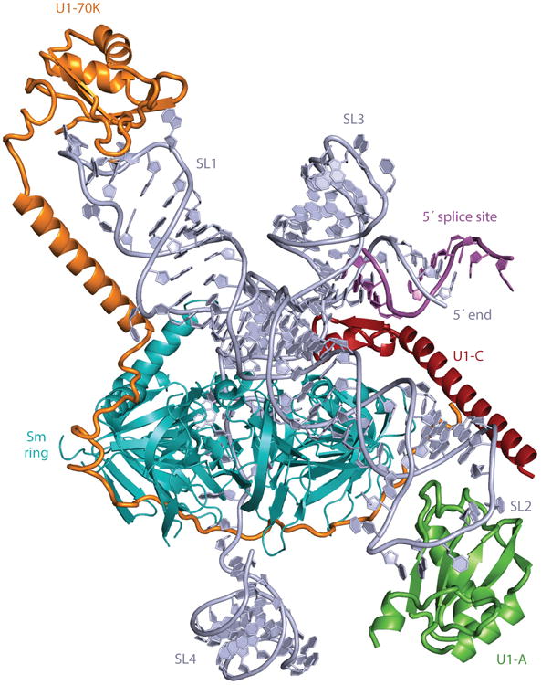
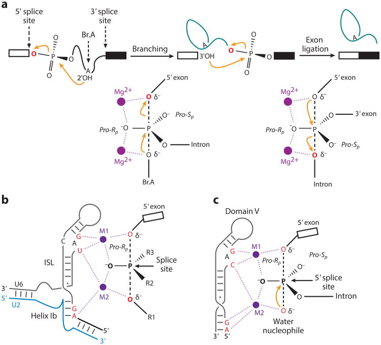
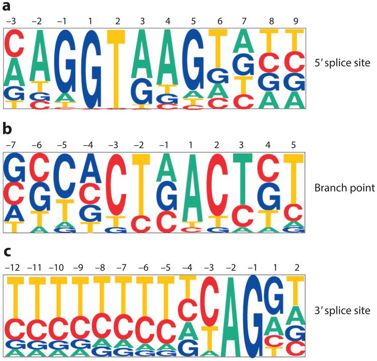
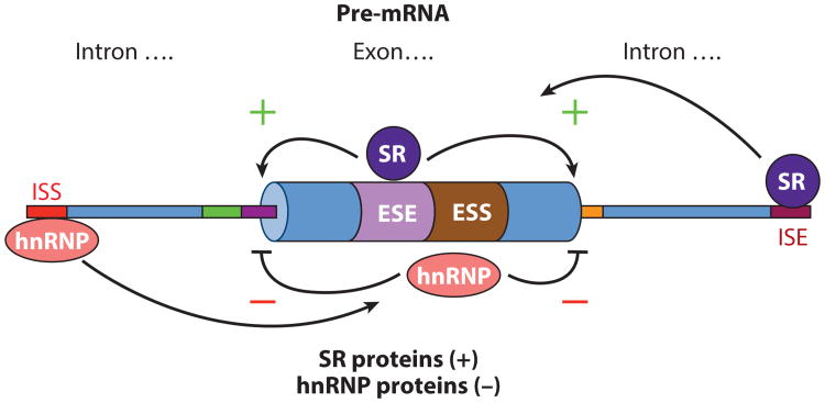
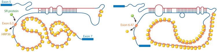
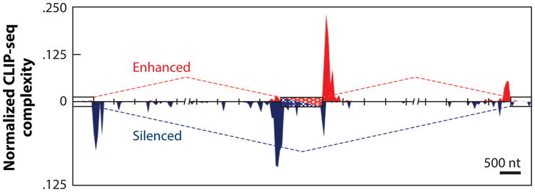
Precursor messenger RNA (pre-mRNA) splicing is a critical step in the posttranscriptional regulation of gene expression, providing significant expansion of the functional proteome of eukaryotic organisms with limited gene numbers. Split eukaryotic genes contain intervening sequences or introns disrupting protein-coding exons, and intron removal occurs by repeated assembly of a large and highly dynamic ribonucleoprotein complex termed the spliceosome, which is composed of five small nuclear ribonucleoprotein particles, U1, U2, U4/U6, and U5. Biochemical studies over the past 10 years have allowed the isolation as well as compositional, functional, and structural analysis of splicing complexes at distinct stages along the spliceosome cycle. The average human gene contains eight exons and seven introns, producing an average of three or more alternatively spliced mRNA isoforms. Recent high-throughput sequencing studies indicate that 100% of human genes produce at least two alternative mRNA isoforms. Mechanisms of alternative splicing include RNA-protein interactions of splicing factors with regulatory sites termed silencers or enhancers, RNA-RNA base-pairing interactions, or chromatin-based effects that can change or determine splicing patterns. Disease-causing mutations can often occur in splice sites near intron borders or in exonic or intronic RNA regulatory silencer or enhancer elements, as well as in genes that encode splicing factors. Together, these studies provide mechanistic insights into how spliceosome assembly, dynamics, and catalysis occur; how alternative splicing is regulated and evolves; and how splicing can be disrupted by cis- and trans-acting mutations leading to disease states. These findings make the spliceosome an attractive new target for small-molecule, antisense, and genome-editing therapeutic interventions.

Keywords: RNA structure; RNA-binding proteins; disease; enhancers; exon; genomics; intron; pre-mRNA splicing; silencers; spliceosome; splicing factors.


Figures

Figure 1
Figure 1
The spliceosome assembly and disassembly cycle, with known structures of individual complexes, as well as the cross-intron assembly and disassembly of the major (U1 and U2) spliceosome. Also depicted is the stepwise interaction of the spliceosomal small nuclear ribonucleoprotein (snRNP) particles (U1, U2, U4, U5, and U6) (colored circles) in the removal of an intron from a precursor messenger RNA (pre-mRNA) containing two exons (blue and purple); non-snRNP proteins are not shown. The spliceosomal complexes that can be resolved biochemically in mammalian splicing extracts are shown. The names of the complexes, as well as the first and second catalytic steps, are indicated. Also shown are the electron microscopy–derived structures of the purified prespliceosome (complex A) (56), the U4/U6.U5 tri-snRNP (249), the precatalytic spliceosome (complex B) (61, 250), and the catalytic step 1 spliceosome (complex C) (52, 60). Abbreviation: SS, splice site. Modified with permission from Reference .
Figure 2
Figure 2
Overview of a model of the complete human U1 small nuclear ribonucleoprotein (snRNP) derived from X-ray crystal structures. Truncated stem loop 2 (SL2) was extended with an A-form RNA helix and, using the crystal structure of the U1A–RNA complex (64), was appended to the extended helix. The internal loop of SL2, consisting of four consecutive non-Watson-Crick base pairs, is in a position to interact with the Sm-B and Sm-D1 proteins. Closely matching images are found in the gallery of negatively stained images of U1 snRNP reported previously (251, 252).
Figure 3
Figure 3
Chemistry of precursor messenger RNA (pre-mRNA) splicing and U2/U6 model showing sites that are sensitive to sulfur substitutions and rescued by thiophilic metal. (a) Reaction scheme (top) and transition state diagrams (bottom) for the two steps of nuclear pre-mRNA splicing. (b) Two-metal model for the RNA catalytic core of the spliceosome. For branching, R1 represents the 29 hydroxyl of the branch adenosine, R2 represents the intron, and R3 represents the pro-Sp oxygen. For exon ligation, R1 represents the 39 oxygen leaving group, R2 represents the pro-Sp oxygen, and R3 represents the 39 exon. (c) Model of group II intron domain V during hydrolysis [PDB 4FAQ (77)]. Throughout, the reactive oxygens are colored red, the pre-mRNA scissile phosphate is depicted in a transition state, and interactions between specific ligands and the reactive oxygens mediated by M1 and M2 are shown as light purple dashed lines. Modified with permission from Reference .
Figure 4
Figure 4
Pictograms of the major U2-dependent intron class consensus splice-site signals. Approximately 20,000 5′ and 3′ splice sites from annotated GenBank files were extracted and aligned as described elsewhere (253, 254). In these pictograms, the size of a letter corresponds to the frequency with which that base is present at each position in a compilation of splice sites. (a) Major class 5′ splice-site consensus sequence. The position labeled 1 is the first nucleotide of the intron, and the position labeled –1 is the last nucleotide of the upstream exon. (b) Major class branch-site consensus. A small database of experimentally confirmed branch sites (166) was used to generate this pictogram. The position labeled 1 is the branch-site residue. (c) Major class 3′ splice-site consensus. The position labeled –1 is the last nucleotide of the intron, and the position labeled 1 is the first nucleotide of the downstream exon. Modified with permission from Reference .
Figure 5
Figure 5
Positive and negative control of precursor messenger RNA (pre-mRNA) splicing by cis-acting intronic and exonic silencers and enhancers. Diagram of a segment of a typical metazoan pre-mRNA with exon and surrounding introns indicated. Intronic and exonic splicing enhancers (ISE, red box; ESE, purple box) and intronic and exonic splicing silencers (ISS, orange box; ESS, brown box) are indicated. Serine–arginine repeat (SR) proteins generally act to promote splicing from nearby splice sites by interacting with splicing enhancers. Heterogeneous nuclear ribonucleoprotein particle (hnRNP) proteins generally act to inhibit splicing from nearby splice sites by interacting with splicing silencers.
Figure 6
Figure 6
RNA–RNA and RNA–protein interactions that regulate mutually exclusive splicing of the Drosophila DSCAM exon 6 exon cluster. A model for the mechanism by which the heterogeneous nuclear ribonucleoprotein (hnRNP) hrp36 prevents the inclusion of multiple DSCAM exon 6 variants. hrp36 (yellow circles) binds to all the exon 6 variants (orange) and represses their inclusion. When the conserved cis-acting RNA selector sequence upstream of a specific exon interacts by RNA–RNA base pairing with the conserved exon 6 cis-acting RNA docking site located upstream of the exon 6 cluster of 48 exons, it results in the derepression of hrp36 on the exon immediately downstream, but not for the other 47 exon 6 variants. In this way, only a single exon 6 variant is included. hrp36 competes with serine–arginine repeat (SR) proteins (green circles) for binding to the exon 6 variants. In the absence of hrp36, these activators can bind to all the exon 6 variants and function to enhance their splicing to other exon 6 exons. Figure modeled on data from Reference .
Figure 7
Figure 7
The RNA map of splicing regulation by the neuronal nova protein. A nova RNA splicing map for cassette exons generated by integrating the high-throughput sequencing cross-linking and immunoprecipitation (HTS-CLIP)/CLIP-sequencing (CLIP-seq) experimental identification of nova-binding sites and splice junction microarray data. The red dashed line (top) indicates enhanced exon inclusion by nova, and red peaks indicate the density and location of HTS-CLIP/CLIP-seq tags. The blue dashed line (bottom) indicates enhanced exon skipping by nova, and blue peaks indicate the density and location of HTS-CLIP/CLIP-seq tags. Modified with permission from Reference .

References

    1. Sharp PA. Split genes and RNA splicing. Cell. 1994;77:805–15. - PubMed
    1. Sharp PA. The discovery of split genes and RNA splicing. Trends Biochem Sci. 2005;30:279–81. - PubMed
    1. Kim E, Magen A, Ast G. Different levels of alternative splicing among eukaryotes. Nucleic Acids Res. 2007;35:125–31. - PMC - PubMed
    1. Kim H, Klein R, Majewski J, Ott J. Estimating rates of alternative splicing in mammals and invertebrates. Nat Genet. 2004;36:915–17. - PubMed
    1. Merkin J, Russell C, Chen P, Burge CB. Evolutionary dynamics of gene and isoform regulation in mammalian tissues. Science. 2012;338:1593–99. - PMC - PubMed

Publication types