

Analysis of the metabolome of Anopheles gambiae mosquito after exposure to Mycobacterium ulcerans
- PMID: 25784490
- PMCID: PMC4363836
- DOI: 10.1038/srep09242
Analysis of the metabolome of Anopheles gambiae mosquito after exposure to Mycobacterium ulcerans
Abstract
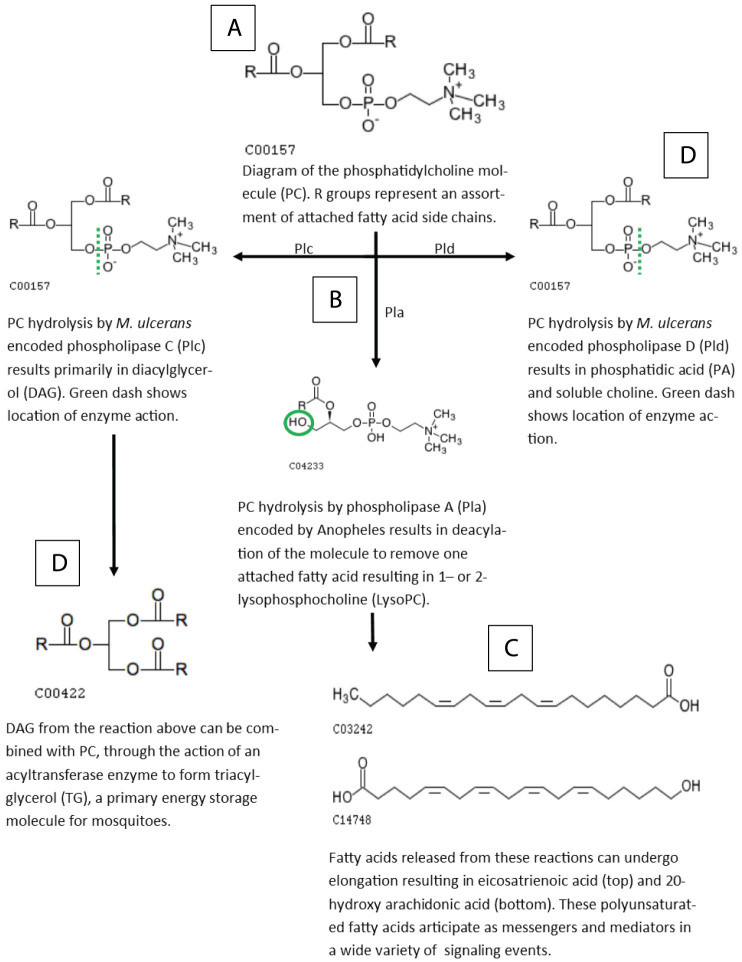
Infection with Mycobacterium ulcerans causes Buruli Ulcer, a neglected tropical disease. Mosquito vectors are suspected to participate in the transmission and environmental maintenance of the bacterium. However, mechanisms and consequences of mosquito contamination by M. ulcerans are not well understood. We evaluated the metabolome of the Anopheles gambiae mosquito to profile the metabolic changes associated with bacterial colonization. Contamination of mosquitoes with live M. ulcerans bacilli results in disruptions to lipid metabolic pathways of the mosquito, specifically the utilization of glycerolipid molecules, an affect that was not observed in mosquitoes exposed to dead M. ulcerans. These results are consistent with aberrations of lipid metabolism described in other mycobacterial infections, implying global host-pathogen interactions shared across diverse saprophytic and pathogenic mycobacterial species. This study implicates features of the bacterium, such as the putative M. ulcerans encoded phospholipase enzyme, which promote virulence, survival, and active adaptation in concert with mosquito development, and provides significant groundwork for enhanced studies of the vector-pathogen interactions using metabolomics profiling. Lastly, metabolic and survival data suggest an interaction which is unlikely to contribute to transmission of M. ulcerans by A. gambiae and more likely to contribute to persistence of M. ulcerans in waters cohabitated by both organisms.
Figures
Similar articles
-
Behavioral interplay between mosquito and mycolactone produced by Mycobacterium ulcerans and bacterial gene expression induced by mosquito proximity.PLoS One. 2023 Aug 3;18(8):e0289768. doi: 10.1371/journal.pone.0289768. eCollection 2023. PLoS One. 2023. PMID: 37535670 Free PMC article.
-
Aquatic snails, passive hosts of Mycobacterium ulcerans.Appl Environ Microbiol. 2004 Oct;70(10):6296-8. doi: 10.1128/AEM.70.10.6296-6298.2004. Appl Environ Microbiol. 2004. PMID: 15466578 Free PMC article.
-
A newly discovered mycobacterial pathogen isolated from laboratory colonies of Xenopus species with lethal infections produces a novel form of mycolactone, the Mycobacterium ulcerans macrolide toxin.Infect Immun. 2005 Jun;73(6):3307-12. doi: 10.1128/IAI.73.6.3307-3312.2005. Infect Immun. 2005. PMID: 15908356 Free PMC article.
-
The genome, evolution and diversity of Mycobacterium ulcerans.Infect Genet Evol. 2012 Apr;12(3):522-9. doi: 10.1016/j.meegid.2012.01.018. Epub 2012 Jan 28. Infect Genet Evol. 2012. PMID: 22306192 Review.
-
Recent advances in leprosy and Buruli ulcer (Mycobacterium ulcerans infection).Curr Opin Infect Dis. 2010 Oct;23(5):445-55. doi: 10.1097/QCO.0b013e32833c2209. Curr Opin Infect Dis. 2010. PMID: 20581668 Review.
Cited by
-
Perplexing Metabolomes in Fungal-Insect Trophic Interactions: A Terra Incognita of Mycobiocontrol Mechanisms.Front Microbiol. 2016 Oct 19;7:1678. doi: 10.3389/fmicb.2016.01678. eCollection 2016. Front Microbiol. 2016. PMID: 27807434 Free PMC article. Review.
-
Metabolomic profiles delineate mycolactone signature in Buruli ulcer disease.Sci Rep. 2015 Dec 4;5:17693. doi: 10.1038/srep17693. Sci Rep. 2015. PMID: 26634444 Free PMC article.
-
Towards a method for cryopreservation of mosquito vectors of human pathogens.Cryobiology. 2021 Apr;99:1-10. doi: 10.1016/j.cryobiol.2021.02.001. Epub 2021 Feb 5. Cryobiology. 2021. PMID: 33556359 Free PMC article. Review.
-
Multiomic interpretation of fungus-infected ant metabolomes during manipulated summit disease.Sci Rep. 2023 Sep 1;13(1):14363. doi: 10.1038/s41598-023-40065-0. Sci Rep. 2023. PMID: 37658067 Free PMC article.
-
Metabolomics of the tick-Borrelia interaction during the nymphal tick blood meal.Sci Rep. 2017 Mar 13;7:44394. doi: 10.1038/srep44394. Sci Rep. 2017. PMID: 28287618 Free PMC article.
References
Publication types
MeSH terms
Substances
Grants and funding
LinkOut - more resources
Full Text Sources
Other Literature Sources
Molecular Biology Databases
