

Specifically targeted delivery of protein to phagocytic macrophages
- PMID: 25784802
- PMCID: PMC4356666
- DOI: 10.2147/IJN.S75950
Specifically targeted delivery of protein to phagocytic macrophages
Abstract
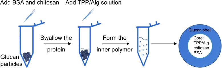
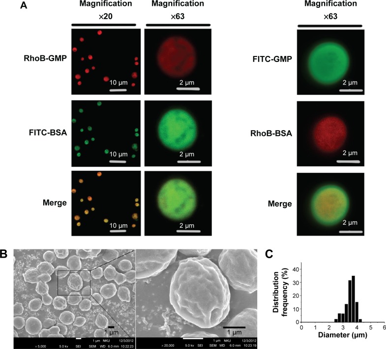
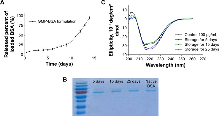
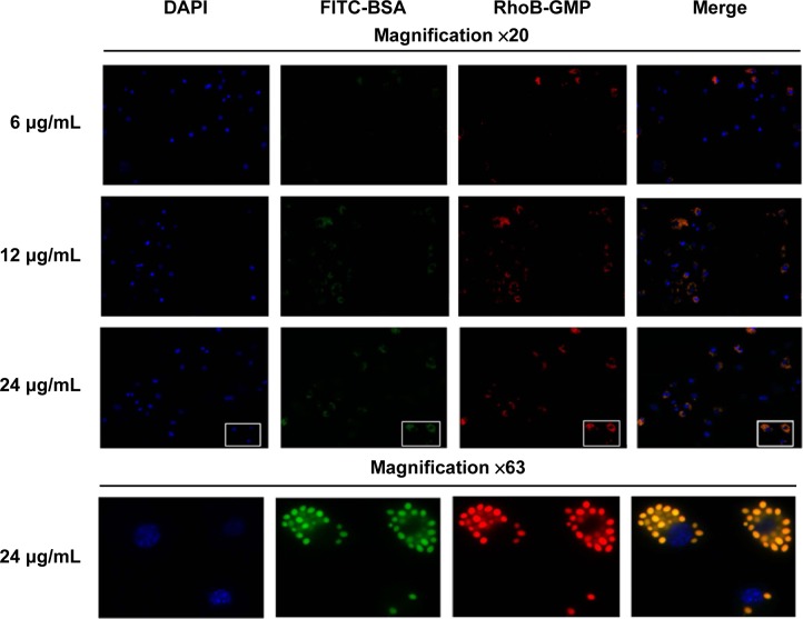
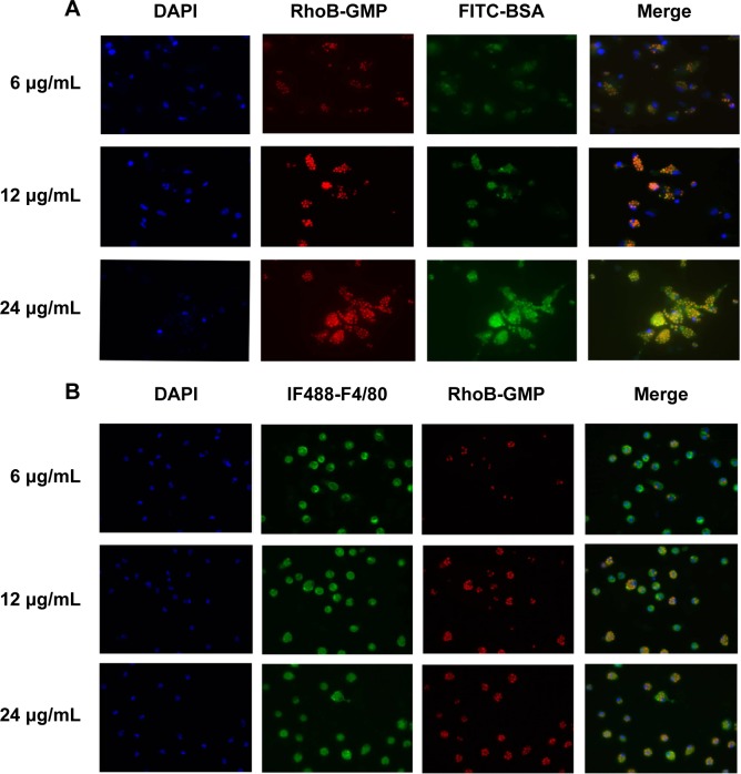
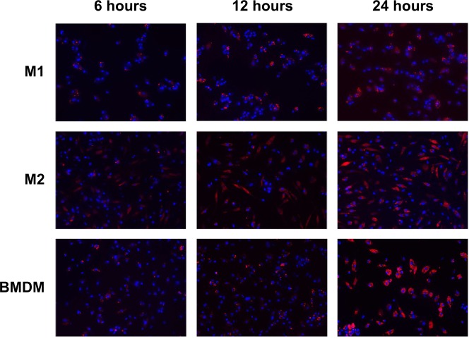
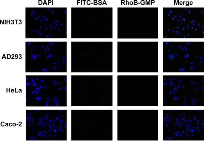
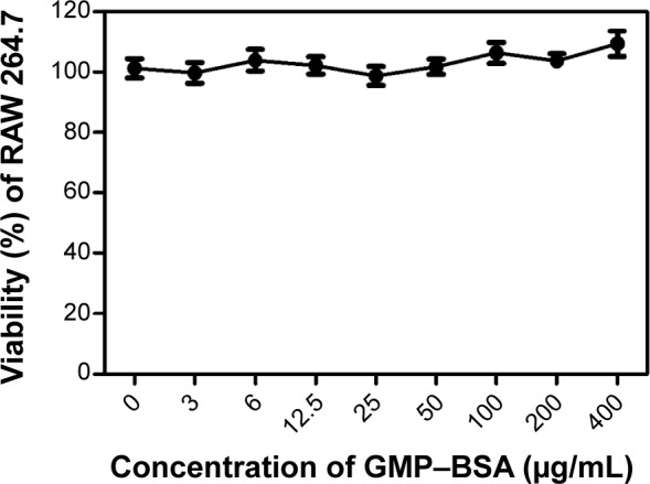
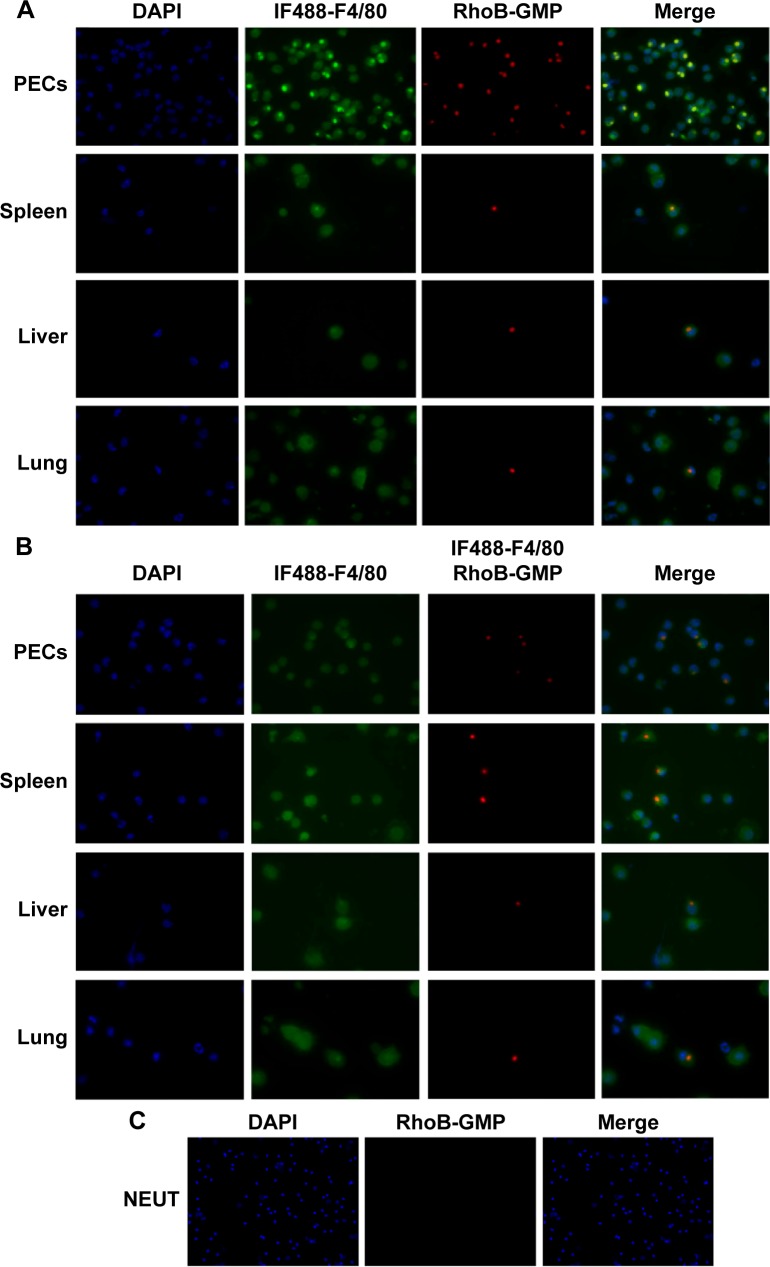
Macrophages play important roles in the pathogenesis of various diseases, and are important potential therapeutic targets. Furthermore, macrophages are key antigen-presenting cells and important in vaccine design. In this study, we report on the novel formulation (bovine serum albumin [BSA]-loaded glucan particles [GMP-BSA]) based on β-glucan particles from cell walls of baker's yeast for the targeted delivery of protein to macrophages. Using this formulation, chitosan, tripolyphosphate, and alginate were used to fabricate colloidal particles with the model protein BSA via electrostatic interactions, which were caged and incorporated BSA very tightly within the β-glucan particle shells. The prepared GMP-BSA exhibited good protein-release behavior and avoided protein leakage. The particles were also highly specific to phagocytic macrophages, such as Raw 264.7 cells, primary bone marrow-derived macrophages, and peritoneal exudate macrophages, whereas the particles were not taken up by nonphagocytic cells, including NIH3T3, AD293, HeLa, and Caco-2. We hypothesize that these tightly encapsulated protein-loaded glucan particles deliver various types of proteins to macrophages with notably high selectivity, and may have broad applications in targeted drug delivery or vaccine design against macrophages.
Keywords: glucan particle; macrophage; targeted drug delivery; vaccine; yeast-cell wall.
Figures
Similar articles
-
Targeted delivery of NK007 to macrophages to treat colitis.J Pharm Sci. 2015 Jul;104(7):2276-84. doi: 10.1002/jps.24473. Epub 2015 May 11. J Pharm Sci. 2015. PMID: 25964181
-
Yeast glucan particles enable intracellular protein delivery in Drosophila without compromising the immune system.Biomater Sci. 2019 Nov 1;7(11):4708-4719. doi: 10.1039/c9bm00539k. Epub 2019 Sep 30. Biomater Sci. 2019. PMID: 31565713
-
Yeast glucan particles: An express train for oral targeted drug delivery systems.Int J Biol Macromol. 2023 Dec 31;253(Pt 5):127131. doi: 10.1016/j.ijbiomac.2023.127131. Epub 2023 Sep 28. Int J Biol Macromol. 2023. PMID: 37776921 Review.
-
Improvement in bioactive protein storage stability and colon-targeted release: a simple double-layer chitosan-based particle.J Microencapsul. 2019 Aug;36(5):474-484. doi: 10.1080/02652048.2019.1646336. Epub 2019 Aug 1. J Microencapsul. 2019. PMID: 31318277
-
β-1,3 Glucan Microparticles & Nanoparticles: Fabrication Methods & Applications in Immunomodulation & Targeted Drug Delivery.Adv Healthc Mater. 2025 May;14(14):e2501006. doi: 10.1002/adhm.202501006. Epub 2025 Apr 29. Adv Healthc Mater. 2025. PMID: 40302314 Free PMC article. Review.
Cited by
-
Activatable biomimetic probe with aggregation-induced emission characteristics for non-invasive monitoring of allograft rejection.Theranostics. 2025 May 30;15(13):6572-6592. doi: 10.7150/thno.110866. eCollection 2025. Theranostics. 2025. PMID: 40521207 Free PMC article.
-
A Potent Micron Neoantigen Tumor Vaccine GP-Neoantigen Induces Robust Antitumor Activity in Multiple Tumor Models.Adv Sci (Weinh). 2022 Aug;9(24):e2201496. doi: 10.1002/advs.202201496. Epub 2022 Jun 16. Adv Sci (Weinh). 2022. PMID: 35712770 Free PMC article.
-
Nanotechnology in Glycomics: Applications in Diagnostics, Therapy, Imaging, and Separation Processes.Med Res Rev. 2017 May;37(3):514-626. doi: 10.1002/med.21420. Epub 2016 Nov 15. Med Res Rev. 2017. PMID: 27859448 Free PMC article. Review.
-
Preparation and characterization of beta-glucan particles containing a payload of nanoembedded rifabutin for enhanced targeted delivery to macrophages.EXCLI J. 2017 Mar 7;16:210-228. doi: 10.17179/excli2016-804. eCollection 2017. EXCLI J. 2017. PMID: 28507467 Free PMC article.
-
Synthesis and Characterization of pH-Sensitive Inulin Conjugate of Isoniazid for Monocyte-Targeted Delivery.Pharmaceutics. 2019 Oct 28;11(11):555. doi: 10.3390/pharmaceutics11110555. Pharmaceutics. 2019. PMID: 31661841 Free PMC article.
References
-
- Gordon S, Martinez FO. Alternative activation of macrophages: mechanism and functions. Immunity. 2010;32(5):593–604. - PubMed
-
- Lebre MC, Tak PP. Macrophage subsets in immune-mediated inflammatory disease: lessons from rheumatoid arthritis, spondyloarthritis, osteoarthritis, Behçet’s disease and gout. Open Arthritis J. 2010;3:18–23.
Publication types
MeSH terms
Substances
LinkOut - more resources
Full Text Sources
Other Literature Sources
