. 2015:2015:502182.
doi: 10.1155/2015/502182. Epub 2015 Feb 16.

Terminalia chebula Fructus Inhibits Migration and Proliferation of Vascular Smooth Muscle Cells and Production of Inflammatory Mediators in RAW 264.7

Affiliations

Terminalia chebula Fructus Inhibits Migration and Proliferation of Vascular Smooth Muscle Cells and Production of Inflammatory Mediators in RAW 264.7

Hyun-Ho Lee et al. Evid Based Complement Alternat Med. 2015.

Abstract

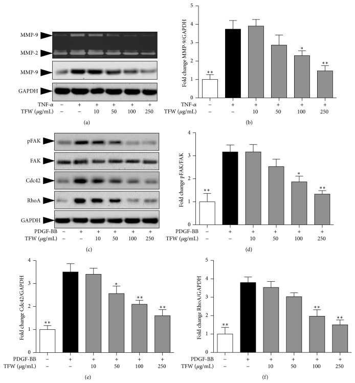
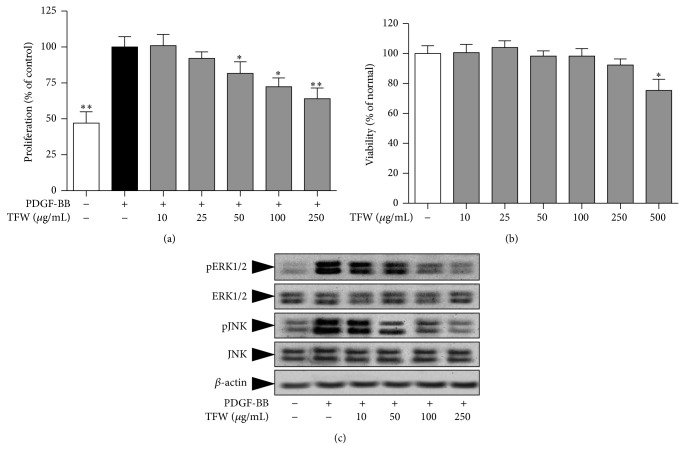
Pathogenesis of atherosclerosis and neointima formation after angioplasty involves vascular smooth muscle cells (VSMCs) migration and proliferation followed by inflammatory responses mediated by recruited macrophages in the neointima. Terminalia chebula is widely used traditional medicine in Asia for its beneficial effects against cancer, diabetes, and bacterial infection. The study was designed to determine whether Terminalia chebula fructus water extract (TFW) suppresses VSMC migration and proliferation and inflammatory mediators production in macrophage (RAW 264.7). Our results showed that TFW possessed strong antioxidative effects in 1,1-diphenyl-2-picryl hydrazyl (DPPH) scavenging and lipid peroxidation assays. In addition, TFW reduced nitric oxide (NO) production, inducible nitric oxide synthase (iNOS), and cyclooxygenase-2 (COX-2) expression in RAW 264.7 cells. Also, TFW inhibited platelet-derived growth factor (PDGF-BB) induced VSMC migration as determined by wound healing and Boyden chamber assays. The antimigratory effect of TFW was due to its inhibitory effect on metalloproteinase-9 (MMP-9) expression, focal adhesion kinase (FAK) activation, and Rho-family of small GTPases (Cdc42 and RhoA) expression in VSMCs. Furthermore, TFW suppressed PDGF-BB induced VSMC proliferation by downregulation of mitogen activated protein kinases (MAPKs) signaling molecules. These results suggest that TFW could be a beneficial resource in the prevention of atherosclerosis.


Figures

Figure 1
Figure 1
DPPH scavenging activity, NO scavenging activity, and metal chelation activity of TFW: (a) DPPH scavenging activity of TFW, gallic acid, and chebulic acid; values are expressed as mean ± standard deviation; n = 5. (b) Chelating activity of TFW, gallic acid, and chebulic acid; values are expressed as mean ± SD, n = 5. (c) Effect of TFW, gallic acid, and chebulic acid on serum lipid peroxidation; values are expressed as mean ± SD; n = 5.
Figure 2
Figure 2
NO production, iNOS, and COX-2 expression in LPS stimulated RAW 264.7. RAW 264.7 cells were pretreated with TFW for 1 h followed by stimulation with LPS for additional 24 h. (a) The cell viability was determined using MTT assay. (b) NO production was measured using Griess reagent. (c) The expression of iNOS and COX-2 was determined by Western blot. β-actin was used for normalization. (d) Normalized quantitative data for iNOS expression. (e) Normalized quantitative data for COX-2 expression. Values are expressed as mean ± SD; n = 4. * P < 0.05 and ** P < 0.01 versus control (LPS alone).
Figure 3
Figure 3
Effect of TFW on VSMC migration. (a) Subconfluent VSMCs were scraped off with a pipette tip to induce wounds and then the cells were treated with TFW for 1 h followed by PDGF-BB treatment. Images of wounded area were captured immediately (time 0) and 6, 12, and 24 h after injury. (b) Finally wound closure was quantified as percentage of the initial wound area that had been recovered with VSMCs. Values are expressed as mean ± SD (n = 5). * P < 0.05 and ** P < 0.01 versus control (PDGF-BB alone); original magnification 200x. (c) The cells were allowed to migrate in the presence of PDGF-BB and the indicated concentrations of TFW for 6 h. After staining with eosin and hematoxylin, the migrated cells were counted (d) in 5 high power fields (200x). Values are expressed as mean ± SD (n = 5). * P < 0.05 and ** P < 0.01 versus control (PDGF-BB alone).
Figure 4
Figure 4
Effect of TFW on MMP-2 and MMP-9 expression. VSMCs were pretreated with TFW for 1 h followed by stimulation with TNF-α for 24 h. (a) The proteolytic activity of MMP-2 and MMP-9 in conditioned medium and cell lysate was measured by gelatin zymography. The protein expression of MMP-9 was determined by Western blot. GAPDH was used for normalization. (b) Normalized quantitative data for MMP-9 protein expression. (c) Serum starved VSMCs were pretreated with indicated concentrations of TFW for 1 h followed by stimulation with PDGF-BB for additional 15 min. The cell lysates were assayed for protein expression of pFAK, FAK, RhoA, and Cdc42. (d) Normalized quantitative data for FAK activation. (e) Normalized quantitative data for Cdc42 expression. (f) Normalized quantitative data for RhoA expression. Values are expressed as mean ± SD; n = 3. * P < 0.05 and ** P < 0.01 versus control (PDGF-BB alone).
Figure 5
Figure 5
Effect of TFW on PDGF-BB induced VSMC proliferation. (a) Serum starved VSMCs were pretreated with indicated concentrations of TFW followed by stimulation with PDGF-BB (20 ng/mL) for 24 h. Cell proliferation was measured by MTT assay and values are expressed as mean ± SD; n = 6. * P < 0.05 and ** P < 0.01 versus control (PDGF-BB alone). (b) Serum starved VSMCs were treated with indicated concentrations of TFW without stimulation for 24 h. Cell viability was measured by MTT assay. (c) Serum starved VSMCs were pretreated with indicated concentrations of TFW for 1 h followed by stimulation with PDGF-BB for additional 15 min. The cell lysates were assayed for protein expression of p-ERK1/2, ERK1/2, pJNK, and JNK. Values are expressed as mean ± SD; n = 6. * P < 0.05 versus normal (untreated).

References

    1. Monraats P. S., Pires N. M. M., Schepers A., et al. Tumor necrosis factor-α plays an important role in restenosis development. The FASEB Journal. 2005;19(14):1998–2004. doi: 10.1096/fj.05-4634com. - DOI - PubMed
    1. Tanizawa S., Ueda M., van der Loos C. M., van der Wal A. C., Becker A. E. Expression of platelet derived growth factor B chain and β receptor in human coronary arteries after percutaneous transluminal coronary angioplasty: an immunohistochemical study. Heart. 1996;75(6):549–556. doi: 10.1136/hrt.75.6.549. - DOI - PMC - PubMed
    1. Ross R. The pathogenesis of atherosclerosis: a perspective for the 1990s. Nature. 1993;362(6423):801–809. doi: 10.1038/362801a0. - DOI - PubMed
    1. Muto A., Fitzgerald T. N., Pimiento J. M., et al. Smooth muscle cell signal transduction: implications of vascular biology for vascular surgeons. Journal of Vascular Surgery. 2007;45(supplement):A15–A24. - PMC - PubMed
    1. Bendeck M. P., Zempo N., Clowes A. W., Galardy R. E., Reidy M. A. Smooth muscle cell migration and matrix metalloproteinase expression after arterial injury in the rat. Circulation Research. 1994;75(3):539–545. doi: 10.1161/01.RES.75.3.539. - DOI - PubMed

LinkOut - more resources