. 2015:2015:593014.
doi: 10.1155/2015/593014. Epub 2015 Feb 16.

Cytotoxic effects of biosynthesized zinc oxide nanoparticles on murine cell lines

Affiliations

Cytotoxic effects of biosynthesized zinc oxide nanoparticles on murine cell lines

Farideh Namvar et al. Evid Based Complement Alternat Med. 2015.

Abstract

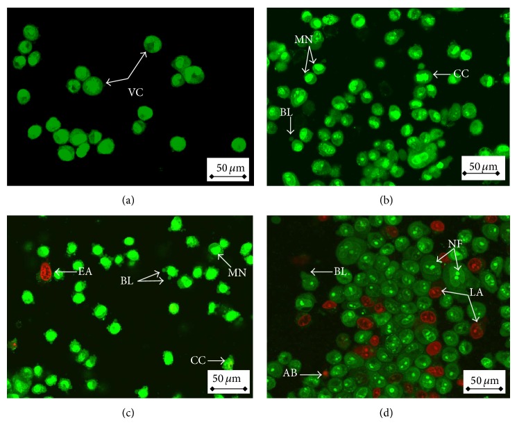
The aim of this study is to evaluate the in vitro cytotoxic activity and cellular effects of previously prepared ZnO-NPs on murine cancer cell lines using brown seaweed (Sargassum muticum) aqueous extract. Treated cancer cells with ZnO-NPs for 72 hours demonstrated various levels of cytotoxicity based on calculated IC50 values using MTT assay as follows: 21.7 ± 1.3 μg/mL (4T1), 17.45 ± 1.1 μg/mL (CRL-1451), 11.75 ± 0.8 μg/mL (CT-26), and 5.6 ± 0.55 μg/mL (WEHI-3B), respectively. On the other hand, ZnO-NPs treatments for 72 hours showed no toxicity against normal mouse fibroblast (3T3) cell line. On the other hand, paclitaxel, which imposed an inhibitory effect on WEHI-3B cells with IC50 of 2.25 ± 0.4, 1.17 ± 0.5, and 1.6 ± 0.09 μg/mL after 24, 48, and 72 hours treatment, respectively, was used as positive control. Furthermore, distinct morphological changes were found by utilizing fluorescent dyes; apoptotic population was increased via flowcytometry, while a cell cycle block and stimulation of apoptotic proteins were also observed. Additionally, the present study showed that the caspase activations contributed to ZnO-NPs triggered apoptotic death in WEHI-3 cells. Thus, the nature of biosynthesis and the therapeutic potential of ZnO-NPs could prepare the way for further research on the design of green synthesis therapeutic agents, particularly in nanomedicine, for the treatment of cancer.


Figures

Figure 1
Figure 1
The aqueous extract of S. muticum (a) before and (b) after synthesis of ZnO-NPs.
Figure 2
Figure 2
(a) FTIR spectra and (b) XRD and (c) UV-visible and (d) TEM image of biosynthesized ZnO-NPs.
Figure 3
Figure 3
(a) Cytotoxic effect of ZnO-NPs on various cancer cells at 72 h of treatment was evaluated through mitochondrial activity using the MTT assay. Each point is the mean value of three replicates. (b) Cytotoxic effects of paclitaxel on WEHI-3B cells at 24, 48, and 72 h of treatment were evaluated through mitochondrial activity using the MTT assay. Each point is the mean value of three replicates. (c) Cytotoxic effects of ZnO-NPs on normal mouse fibroblast cell line (3T3) at 72 h of treatment were evaluated with MTT assay. Each point is the mean value of three replicates.
Figure 4
Figure 4
Fluorescent micrograph of AO/PI double stained WEHI-3B cells that was treated with ZnO-NPs. (a) Untreated cells showing normal cell structure. (b) Early apoptotic cells after 24 h treatment showing membrane blebbing and chromatin condensation. (c) Blebbing and nuclear margination after 48 h treatment. (d) DNA fragmentation and apoptotic body formation after 72 h treatment. VC: viable cells; EA: early apoptotic cells; CC: chromatin condensation; BL: blebbing; MN: marginated nucleus; FN: fragmented nucleus; LA: late apoptotic cells; and AB: apoptotic body.
Figure 5
Figure 5
Flow cytometric analysis of apoptosis induction by ZnO-NPs in WEHI-3B cells after staining with FITC-conjugated annexin V and PI. (a1)–(c1) untreated (control) WEHI-3B cells at 12, 24, and 48 h incubation, respectively. (a2)–(c2) effects of 12, 24, and 48 h ZnO-NPs treatment, respectively.
Figure 6
Figure 6
Cell cycle analysis of WEHI-3B cells treated with ZnO-NPs after staining with PI. (a1)–(c1) untreated WEHI-3B cells for 24, 48, and 72 h, respectively. (a2)–(c2) the effects of 24, 48, and 72 h, respectively, relative to exposure of WEHI-3B cells to ZnO-NPs. G0/G1, G2/M, and S indicate the cell phase, and Sub-G0-G1 refers to the apoptotic cells.
Figure 7
Figure 7
Effect of ZnO-NPs treatment on WEHI-3B cell caspase-3 and caspase-9. The values are mean % ± SD of three independent experiments. Significant differences (P < 0.05) between treated and control groups for caspase-3 and caspase-9 were found.
Figure 8
Figure 8
(a) Protein expression in treated WEHI-3B with ZnO-NPs for 24, 48, and 72 h observed by Western blotting assay. (b) Western blot transcription analysis of treated WEHI-3B with ZnO-NPs for 24, 48, and 72 h. Data analyzed using post hoc comparison of one-way ANOVA using Tukey's b test. The results showed significant (P < 0.05) Bax protein expressions and Bcl-2 protein suppression in all treated groups.

References

    1. Maeng J. H., Lee D.-H., Jung K. H., et al. Multifunctional doxorubicin loaded superparamagnetic iron oxide nanoparticles for chemotherapy and magnetic resonance imaging in liver cancer. Biomaterials. 2010;31(18):4995–5006. doi: 10.1016/j.biomaterials.2010.02.068. - DOI - PubMed
    1. Salata O. V. Applications of nanoparticles in biology and medicine. Journal of Nanobiotechnology. 2004;2, article 3 doi: 10.1186/1477-3155-2-3. - DOI - PMC - PubMed
    1. Kim P. S., Djazayeri S., Zeineldin R. Novel nanotechnology approaches to diagnosis and therapy of ovarian cancer. Gynecologic Oncology. 2011;120(3):393–403. doi: 10.1016/j.ygyno.2010.11.029. - DOI - PubMed
    1. Wang L.-S., Chuang M.-C., Ho J.-A. A. Nanotheranostics—a review of recent publications. International Journal of Nanomedicine. 2012;7:4679–4695. doi: 10.2147/ijn.s33065. - DOI - PMC - PubMed
    1. Jha A. K., Prasad K., Kulkarni A. R. Plant system: nature's nanofactory. Colloids and Surfaces B: Biointerfaces. 2009;73(2):219–223. doi: 10.1016/j.colsurfb.2009.05.018. - DOI - PubMed

LinkOut - more resources