. 2015 Mar 7:12:2.
doi: 10.1186/s12979-015-0029-9. eCollection 2015.

Blood-brain barrier dysfunction developed during normal aging is associated with inflammation and loss of tight junctions but not with leukocyte recruitment

Affiliations

Blood-brain barrier dysfunction developed during normal aging is associated with inflammation and loss of tight junctions but not with leukocyte recruitment

Mina Elahy et al. Immun Ageing. .

Abstract

Background: Functional loss of blood-brain barrier (BBB) is suggested to be pivotal to pathogenesis and pathology of vascular-based neurodegenerative disorders such as Alzheimer's disease. We recently reported in wild-type mice maintained on standard diets, progressive deterioration of capillary function with aging concomitant with heightened neuroinflammation. However, the mice used in this study were relatively young (12 months of age) and potential mechanisms for loss of capillary integrity were not investigated per se. The current study therefore extended the previous finding to investigate the effect of aging on BBB integrity in aged mice at 24 months and its potential underlying molecular mechanisms.

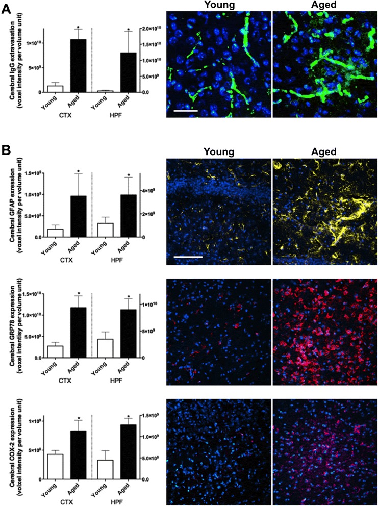
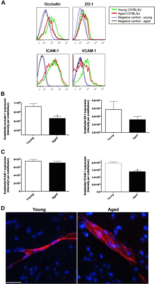
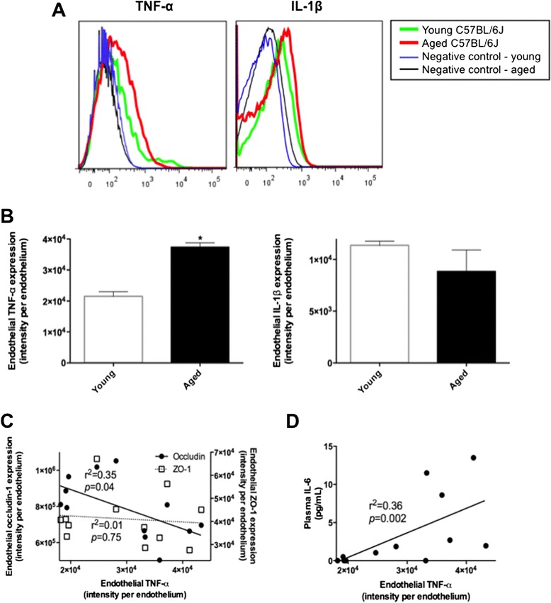
Results: Immunomicroscopy analyses confirmed significantly increased capillary permeability with heightened neuroinflammation in naturally aged 24-month old mice compared to young control at 3 months of age. Aged mice showed significant attenuation in the expression of BBB tight junction proteins, occludin-1 and to lesser extent ZO-1 compared to young mice. In addition, TNF-α in cerebral endothelial cells of aged mice was significantly elevated compared to controls and this was associated with heightened peripheral inflammation. The expression of ICAM-1 and VCAM-1 remained unelevated, and no sign of leukocyte recruitment was observed in aged mice.

Conclusion: The BBB breakdown that occurs during ordinary aging is associated with inflammation and disruption of tight junction complex assembly but not through leukocyte trafficking.

Keywords: Aging; Blood–brain barrier; Inflammation; Leukocyte infiltration; Neurodegenerative disorder; Neuroinflammation; Tight junction complex.


Figures

Figure 1
Figure 1
Blood–brain barrier integrity and neuroinflammation. (A) The integrity of BBB was assessed by measuring the blood-to-brain extravasation of IgG with semi-quantitative confocal microscopy in the cortex (CTX) and hippocampal formation (HPF) of 3 months old young mice and 24 months old aged mice. The voxel intensity of protein of interest is expressed as per volume unit. Asterisks indicate statistical significance assessed with two-tailed t-test (p < 0.05, n = 6). Representative immunomicrographs are also shown (green = IgG, blue = DAPI, scale bar = 20 μm). (B) The astroglial activation, neuronal ER stress and inflammation were assessed by measuring the expression of GFAP, GRP78 and COX-2 respectively. Representative immunomicrographs are shown (yellow = GFAP, red = GRP78, magenta = COX-2, blue = DAPI, scale bar = 50 μm).
Figure 2
Figure 2
Cerebrovascular tight junction proteins and leukocyte recruitment. BBB tight junction assembly was assessed by measuring endothelial cell expressions of occludin-1 and ZO-1 with flow cytometry in the brains of 3 months old young mice and 24 months old aged mice. Example plots are in (A) and the fluorescent intensity of protein of interest is expressed as per endothelial cell (B). Asterisks indicate statistical significance assessed with two-tailed t-test (p < 0.05, n = 6). (C) Endothelial expression of adhesion proteins, ICAM-1 and VCAM-1, was measured with flow cytometry. (D) The infiltration of leukocytes across the BBB was assessed with immunomicroscopy staining of CD45 immunoreactivity within the perivascular region of entire cortex and hippocampal formation. Representative images are shown. Scale bar indicates 20 μm.
Figure 3
Figure 3
Blood–brain barrier endothelial levels of pro-inflammatory cytokines. The intracellular levels of TNF-α and IL-1β in cerebrovascular endothelial cells were measured with flow cytometry in the brains of 3 months old young mice and 24 months old aged mice. Example plots in (A) and mean of n = 6 in (B). Asterisks indicate statistical significance assessed with two-tailed t-test (p < 0.05, n = 6). (C) The causal association of attenuated BBB tight junction proteins, occludin-1 or ZO-1, with endothelial TNF-α was analyzed with Pearson’s correlation coefficient (n = 12). (D) The association between the cerebrovascular TNF-α and peripheral IL-6 was determined with Pearson’s correlation coefficient (n = 12).

References

    1. Zlokovic BV. Neurovascular pathways to neurodegeneration in Alzheimer's disease and other disorders. Nat Rev Neurosci. 2011;12:723–38. - PMC - PubMed
    1. Zlokovic BV. The blood–brain barrier in health and chronic neurodegenerative disorders. Neuron. 2008;57:178–201. doi: 10.1016/j.neuron.2008.01.003. - DOI - PubMed
    1. Marques F, Sousa JC, Sousa N, Palha JA. Blood–brain-barriers in aging and in Alzheimer's disease. Mol Neurodegener. 2013;8:38. doi: 10.1186/1750-1326-8-38. - DOI - PMC - PubMed
    1. Farrall AJ, Wardlaw JM. Blood–brain barrier: ageing and microvascular disease–systematic review and meta-analysis. Neurobiol Aging. 2009;30:337–52. doi: 10.1016/j.neurobiolaging.2007.07.015. - DOI - PubMed
    1. De Reuck JL. Histopathological stainings and definitions of vascular disruptions in the elderly brain. Exp Gerontol. 2012;47:834–7. doi: 10.1016/j.exger.2012.03.012. - DOI - PubMed

LinkOut - more resources