. 2015 Feb 24;9(1):014126.
doi: 10.1063/1.4913648. eCollection 2015 Jan.

Enhanced fluorescence emitted by microdroplets containing organic dye emulsions

Affiliations

Enhanced fluorescence emitted by microdroplets containing organic dye emulsions

M Boni et al. Biomicrofluidics. .

Abstract

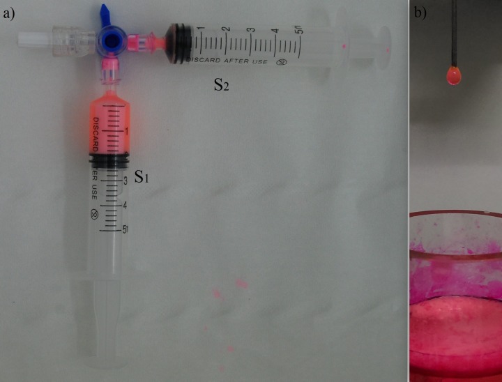
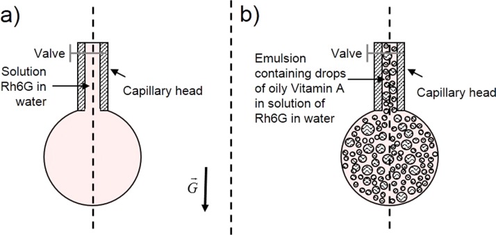
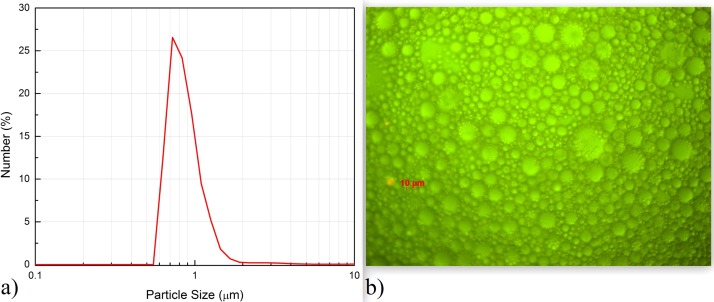
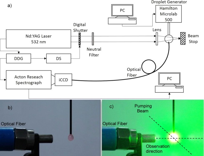
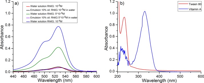
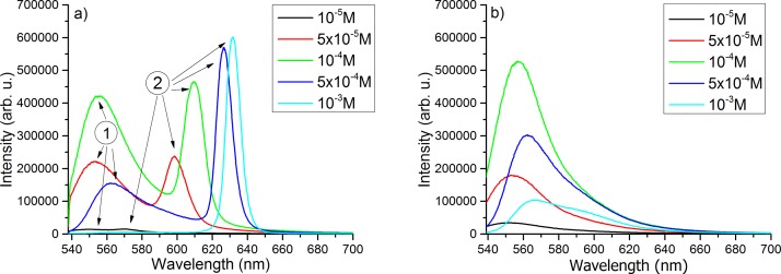
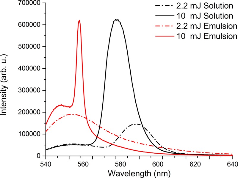
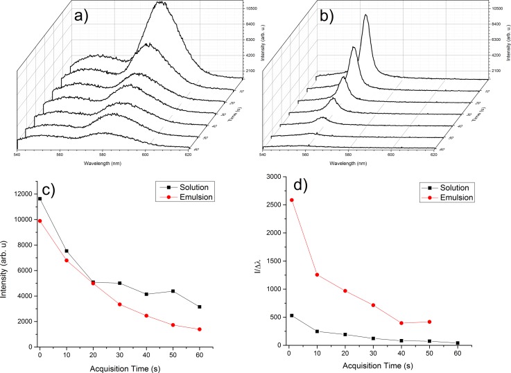
In this paper, laser beam resonant interaction with pendant microdroplets that are seeded with a laser dye (Rhodamine 6G (Rh6G)) water solution or oily Vitamin A emulsion with Rhodamine 6G solution in water is investigated through fluorescence spectra analysis. The excitation is made with the second harmonic generated beam emitted by a pulsed Nd:YAG laser system at 532 nm. The pendant microdroplets containing emulsion exhibit an enhanced fluorescence signal. This effect can be explained as being due to the scattering of light by the sub-micrometric drops of oily Vitamin A in emulsion and by the spherical geometry of the pendant droplet. The droplet acts as an optical resonator amplifying the fluorescence signal with the possibility of producing lasing effect. Here, we also investigate how Rhodamine 6G concentration, pumping laser beam energies and number of pumping laser pulses influence the fluorescence behavior. The results can be useful in optical imaging, since they can lead to the use of smaller quantities of fluorescent dyes to obtain results with the same quality.


Figures

FIG. 1.
FIG. 1.
DSS used to generate emulsion. (a) Emulsion accumulated in syringe S1 at the end of pumping sequence. (b) Pendant droplet containing emulsion extracted from a crucible where S1 was emptied.
FIG. 2.
FIG. 2.
Cartoon depicting pendant droplets containing (a) solution of Rh6G in water and (b) emulsion of oily Vitamin A and Rh6G water solution.
FIG. 3.
FIG. 3.
Oil microdroplets distribution in emulsion. (a) Oil droplets size distribution obtained by light scattering using Malvern Mastersizer E2000 system. (b) Image of the emulsion structure recorded with a Zeiss Optical Microscope (Imager. Z1m) equipped with axio Cam MRc 5 (HR).
FIG. 4.
FIG. 4.
Scheme of the experimental set-up. (a) Set-up structure. (b) Pendant droplet of emulsion prior exposure to pumping beam and (c) during exposure.
FIG. 5.
FIG. 5.
Absorption spectra of the compounds used in the experiments. (a) Absorption spectra of Rh6G solutions in water and of emulsions produced by mixing water Rh6G solution and Vitamin A. (b) Absorption spectra of Tween 80 and Vitamin A.
FIG. 6.
FIG. 6.
Laser induced fluorescence emitted by pendant droplets. (a) Lasing and fluorescence spectra obtained from droplet of Rh6G solution in distilled water. (b) Fluorescence spectra obtained in a pendant droplet containing a mixture of Rh6G solution and Oily Vitamin A.
FIG. 7.
FIG. 7.
Lasing and fluorescence spectra obtained in pendant droplet containing an emulsion of Rh6G in water at 5 × 10−5M and oily Vitamin A for two pumping energies 2.2 mJ and 10 mJ.
FIG. 8.
FIG. 8.
Time evolution of emission spectra obtained for pendant droplets exposed 1 min (600 pumping pulses). Energy per pumping pulse 10 mJ; each spectrum is an average of the fluorescence emission measured for 10 pulses. (a) Pendant droplet containing Rh6G solution (5 × 10−5M) in distilled water. (b) Pendant droplet containing emulsion of Rh6G solution in water (5 × 10−5M) and Oily Vitamin A. (c) Comparison of lasing intensity decreasing in time for both cases. (d) Lasing intensity/lasing bandwidth ratio function of data acquisition time.

References

    1. Fort E. and Grésillon S., “ Surface enhanced fluorescence,” J. Phys. Appl. Phys. 41, 013001 (2008).10.1088/0022-3727/41/1/013001 - DOI
    1. Kolmakov K.et al., “ Red-emitting rhodamine dyes for fluorescence microscopy and nanoscopy,” Chem. Weinh. Bergstr. Ger. 16, 158–166 (2010).10.1002/chem.200902309 - DOI - PubMed
    1. Klymchenko A. S.et al., “ Highly lipophilic fluorescent dyes in nano-emulsions: Towards bright non-leaking nano-droplets,” RSC Adv. 2, 11876–11886 (2012).10.1039/c2ra21544f - DOI - PMC - PubMed
    1. Saetzler R. K.et al., “ Intravital fluorescence microscopy: Impact of light-induced phototoxicity on adhesion of fluorescently labeled leukocytes,” J. Histochem. Cytochem. Off. J. Histochem. Soc. 45, 505–513 (1997).10.1177/002215549704500403 - DOI - PubMed
    1. van de Hulst H. C., Light Scattering by Small Particles ( Dover Publications, 1981).