Peripheral prism glasses: effects of moving and stationary backgrounds
- PMID: 25785533
- PMCID: PMC4424073
- DOI: 10.1097/OPX.0000000000000552
Peripheral prism glasses: effects of moving and stationary backgrounds
Abstract
Purpose: Unilateral peripheral prisms for homonymous hemianopia (HH) expand the visual field through peripheral binocular visual confusion, a stimulus for binocular rivalry that could lead to reduced predominance and partial suppression of the prism image, thereby limiting device functionality. Using natural-scene images and motion videos, we evaluated whether detection was reduced in binocular compared with monocular viewing.
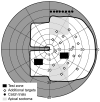
Methods: Detection rates of nine participants with HH or quadranopia and normal binocularity wearing peripheral prisms were determined for static checkerboard perimetry targets briefly presented in the prism expansion area and the seeing hemifield. Perimetry was conducted under monocular and binocular viewing with targets presented over videos of real-world driving scenes and still frame images derived from those videos.
Results: With unilateral prisms, detection rates in the prism expansion area were significantly lower in binocular than in monocular (prism eye) viewing on the motion background (medians, 13 and 58%, respectively, p = 0.008) but not the still frame background (medians, 63 and 68%, p = 0.123). When the stimulus for binocular rivalry was reduced by fitting prisms bilaterally in one HH and one normally sighted subject with simulated HH, prism-area detection rates on the motion background were not significantly different (p > 0.6) in binocular and monocular viewing.
Conclusions: Conflicting binocular motion appears to be a stimulus for reduced predominance of the prism image in binocular viewing when using unilateral peripheral prisms. However, the effect was only found for relatively small targets. Further testing is needed to determine the extent to which this phenomenon might affect the functionality of unilateral peripheral prisms in more real-world situations.
Figures


Similar articles
-
Peripheral prism glasses: effects of dominance, suppression, and background.Optom Vis Sci. 2012 Sep;89(9):1343-52. doi: 10.1097/OPX.0b013e3182678d99. Optom Vis Sci. 2012. PMID: 22885783 Free PMC article.
-
Binocular rivalry with peripheral prisms used for hemianopia rehabilitation.Ophthalmic Physiol Opt. 2014 Sep;34(5):573-9. doi: 10.1111/opo.12143. Ophthalmic Physiol Opt. 2014. PMID: 25160892
-
No Useful Field Expansion with Full-field Prisms.Optom Vis Sci. 2018 Sep;95(9):805-813. doi: 10.1097/OPX.0000000000001271. Optom Vis Sci. 2018. PMID: 30169356 Free PMC article.
-
Monovision.J Am Optom Assoc. 1990 Nov;61(11):820-6. J Am Optom Assoc. 1990. PMID: 2081823 Review.
-
Review: Binocular double vision in the presence of visual field loss.J Vis. 2024 Jun 3;24(6):13. doi: 10.1167/jov.24.6.13. J Vis. 2024. PMID: 38899959 Free PMC article. Review.
Cited by
-
Multiplexing Prisms for Field Expansion.Optom Vis Sci. 2017 Aug;94(8):817-829. doi: 10.1097/OPX.0000000000001102. Optom Vis Sci. 2017. PMID: 28727615 Free PMC article.
-
The effect of visual rivalry in peripheral head-mounted displays on mobility.Sci Rep. 2023 Nov 18;13(1):20199. doi: 10.1038/s41598-023-47427-8. Sci Rep. 2023. PMID: 37980436 Free PMC article.
-
Binocular see-through configuration and eye movement attenuate visual rivalry in peripheral wearable displays.Proc SPIE Int Soc Opt Eng. 2023 Jan-Feb;12449:124490T. doi: 10.1117/12.2648481. Epub 2023 Mar 16. Proc SPIE Int Soc Opt Eng. 2023. PMID: 36970500 Free PMC article.
-
Hazard detection with a monocular bioptic telescope.Ophthalmic Physiol Opt. 2015 Sep;35(5):530-9. doi: 10.1111/opo.12232. Ophthalmic Physiol Opt. 2015. PMID: 26303448 Free PMC article.
-
2017 Charles F. Prentice Award Lecture: Peripheral Prisms for Visual Field Expansion: A Translational Journey.Optom Vis Sci. 2020 Oct;97(10):833-846. doi: 10.1097/OPX.0000000000001590. Optom Vis Sci. 2020. PMID: 33055514 Free PMC article.
References
-
- Zhang X, Kedar S, Lynn MJ, Newman NJ, Biousse V. Homonymous hemianopias: clinical-anatomic correlations in 904 cases. Neurology. 2006;66:906–10. - PubMed
-
- Chen CS, Lee AW, Clarke G, Hayes A, George S, Vincent R, Thompson A, Centrella L, Johnson K, Daly A, Crotty M. Vision-related quality of life in patients with complete homonymous hemianopia post stroke. Top Stroke Rehabil. 2009;16:445–53. - PubMed
-
- Papageorgiou E, Hardiess G, Schaeffel F, Wiethoelter H, Karnath HO, Mallot H, Schoenfisch B, Schiefer U. Assessment of vision-related quality of life in patients with homonymous visual field defects. Graefes Arch Clin Exp Ophthalmol. 2007;245:1749–58. - PubMed
Publication types
MeSH terms
Grants and funding
LinkOut - more resources
Full Text Sources
Medical

