. 2015 Apr 15;137(14):4771-7.
doi: 10.1021/jacs.5b00315. Epub 2015 Apr 1.

Design of a highly selective quenched activity-based probe and its application in dual color imaging studies of cathepsin S activity localization

Affiliations

Design of a highly selective quenched activity-based probe and its application in dual color imaging studies of cathepsin S activity localization

Kristina Oresic Bender et al. J Am Chem Soc. .

Abstract

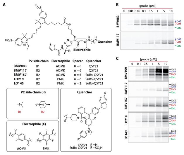
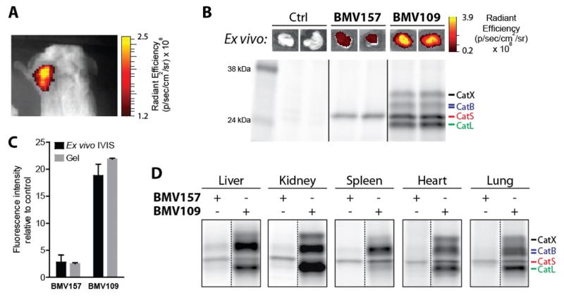
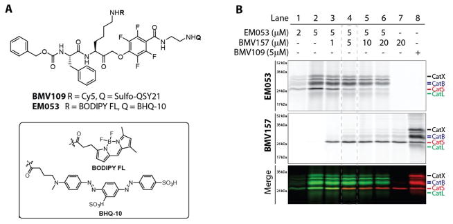
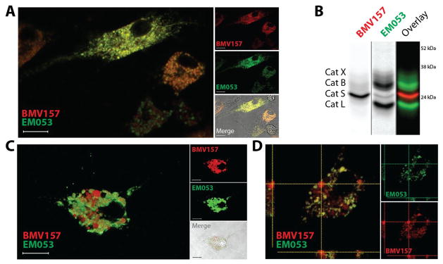
The cysteine cathepsins are a group of 11 proteases whose function was originally believed to be the degradation of endocytosed material with a high degree of redundancy. However, it has become clear that these enzymes are also important regulators of both health and disease. Thus, selective tools that can discriminate between members of this highly related class of enzymes will be critical to further delineate the unique biological functions of individual cathepsins. Here we present the design and synthesis of a near-infrared quenched activity-based probe (qABP) that selectively targets cathepsin S which is highly expressed in immune cells. Importantly, this high degree of selectivity is retained both in vitro and in vivo. In combination with a new green-fluorescent pan-reactive cysteine cathepsin qABP we performed dual color labeling studies in bone marrow-derived immune cells and identified vesicles containing exclusively cathepsin S activity. This observation demonstrates the value of our complementary cathepsin probes and provides evidence for the existence of specific localization of cathepsin S activity in dendritic cells.


Conflict of interest statement

The authors declare no competing financial interest.

Figures

Figure 1
Figure 1. The development of a cathepsin S selective qABP
A) Structures of the qABP BMV083 and the novel probes synthesized and characterized in this work. B) Concentration-dependent labeling profile of BMV083 compared to BMV117 in live RAW cells. C) Concentration-dependent labeling profile of BMV109 compared to BMV117, BMV157, LO219 and LO143 in live RAW cells.
Figure 2
Figure 2. In vivo characterization of the cathepsin S selective qABP BMV157 in a syngeneic orthotopic mouse breast cancer model
A) Noninvasive optical imaging of 4T1 tumor-bearing Balb/c mice injected with BMV157. B) Ex vivo tumor fluorescence (top panel) and in vivo fluorescently labeled tumor-associated proteins visualized after SDS-PAGE by in-gel fluorescence scanning (lower panel) of tumor-bearing mice treated with vehicle, BMV157 or BMV109. C) Quantification of tumor cysteine cathepsin labeling intensity of the ex vivo IVIS measurements and fluorescence SDS-PAGE. D) In vivo tissue specific labeling profile of BMV157 compared to the pan-reactive qABP BMV109 visualized after SDS-PAGE by in-gel fluorescence scanning.
Figure 3
Figure 3. Complementary cysteine cathepsin labeling
A) Structures of the pan-reactive cysteine cathepsin qABPs BMV109 and EM053. B) Labeling profile and simultaneous multi-color labeling of cathepsin S with the near infra-red labeled BMV157 and the green pan-reactive EM053 in living human monocyte-derived dendritic cells.
Figure 4
Figure 4. Live cell co-localization of cathepsin S and other cysteine cathepsin activity using the novel complementary set of qABPs
A) Live cell confocal microscopy and B) biochemical characterization of BMV157 (5 μM) and EM053 (5 μM) co-treated mouse bone marrow-derived macrophages. C, D) Confocal microscopy of mouse bone marrow-derived dendritic cells exposed to BMV157 (5 μM) and EM053 (5 μM). Three-dimensional reconstruction of confocal microscopy images of BMV157 labeled cathepsin S (red) and pan-cathepsin labeling with EM053 (green) (C). Orthogonal projection of the confocal images showing co-localization and lack thereof of BMV157 and EM053 labeled cathepsins (D). Scale bar 10μm.

Similar articles

Cited by

References

    1. Rawlings ND, Barrett AJ. Nucleic Acids Res. 1999;27:325–331. - PMC - PubMed
    1. Turk B, Turk D, Turk V. EMBO J. 2012;31:1630–1643. - PMC - PubMed
    1. Conus S, Simon HU. Swiss Med Wkly. 2010;140:w13042. - PubMed
    1. Driessen C, Bryant RA, Lennon-Dumenil AM, Villadangos JA, Bryant PW, Shi GP, Chapman HA, Ploegh HL. J Cell Biol. 1999;147:775–790. - PMC - PubMed
    1. Beers C, Burich A, Kleijmeer MJ, Griffith JM, Wong P, Rudensky AY. J Immunol. 2005;174:1205–1212. - PubMed

Publication types