. 2015 Apr 7;54(13):2249-2261.
doi: 10.1021/acs.biochem.5b00061. Epub 2015 Mar 24.

Self-assembly of a nine-residue amyloid-forming peptide fragment of SARS corona virus E-protein: mechanism of self aggregation and amyloid-inhibition of hIAPP

Affiliations

Self-assembly of a nine-residue amyloid-forming peptide fragment of SARS corona virus E-protein: mechanism of self aggregation and amyloid-inhibition of hIAPP

Anirban Ghosh et al. Biochemistry. .

Abstract

Molecular self-assembly, a phenomenon widely observed in nature, has been exploited through organic molecules, proteins, DNA, and peptides to study complex biological systems. These self-assembly systems may also be used in understanding the molecular and structural biology which can inspire the design and synthesis of increasingly complex biomaterials. Specifically, use of these building blocks to investigate protein folding and misfolding has been of particular value since it can provide tremendous insights into peptide aggregation related to a variety of protein misfolding diseases, or amyloid diseases (e.g., Alzheimer's disease, Parkinson's disease, type-II diabetes). Herein, the self-assembly of TK9, a nine-residue peptide of the extra membrane C-terminal tail of the SARS corona virus envelope, and its variants were characterized through biophysical, spectroscopic, and simulated studies, and it was confirmed that the structure of these peptides influences their aggregation propensity, hence, mimicking amyloid proteins. TK9, which forms a beta-sheet rich fibril, contains a key sequence motif that may be critical for beta-sheet formation, thus making it an interesting system to study amyloid fibrillation. TK9 aggregates were further examined through simulations to evaluate the possible intra- and interpeptide interactions at the molecular level. These self-assembly peptides can also serve as amyloid inhibitors through hydrophobic and electrophilic recognition interactions. Our results show that TK9 inhibits the fibrillation of hIAPP, a 37 amino acid peptide implicated in the pathology of type-II diabetes. Thus, biophysical and NMR experimental results have revealed a molecular level understanding of peptide folding events, as well as the inhibition of amyloid-protein aggregation are reported.


Figures
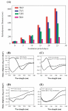
Figure 1
Figure 1
(A) Dynamic light scattering (DLS) plot for TK9 (red), TY5 (blue), YR5 (green) and SK4 (pink);the intensity vs. size distribution bar diagram are plotted at different time intervals. (B – E) Circular Dichroism (CD) plots of secondary structure of TK9 and its fragments measured after freshly preparation and incubation with 15 days.
Figure 2
Figure 2
Transmission and scanning electron micrographs showing fibrillar nanostructure morphology for TK9 (A and C) and TY5 (B and D), respectively.
Figure 3
Figure 3
(A) Tyrosine fluorescence of TK9 (red), TY5 (black) and YR5 (blue) monitoring increase in Tyr fluorescence signal as peptide aggregates. (B) Thioflavin T (ThT) fluorescence assay measuring β-sheet rich fibril formation of freshly dissolved TK9 (black) and after 16 days of TK9 incubation (red).
Figure 4
Figure 4
(A) Amide proton chemical shift regions of 1HNMR spectra of TK9 (A), TY5 (B), YR5 (C) and SK4 (D); (E) average decrease in peak intensity for each peptide. Spectra obtained after 0, 15 and 25 days incubation period are overlaid.
Figure 5
Figure 5
The residue wise relaxation profile for freshly prepared TK9 (blue) and 25 days incubated TK9 (red). (A) Longitudinal relaxation rate (R1) and (B) transverse relaxation rate (R2) are plotted for each residue of TK9.
Figure 6
Figure 6
(A) Pairwise hydrogen bond analysis between any two monomers. Cumulative % H bond occupancy is calculated for respective residue pair in each peptide. (B) Percentage of H-bond occupancy of respective residue in each monomer was calculated to understand the amino acid-solvent interactions. Red bar represents the assembled and blue bar represents unassembled aggregation of each monomer of TK9. (C) Total number of hydrophobic interactions for respective residue pair is calculated for all the peptides forming cluster. (D) Oligomeric structure of TK9 is stabilized by hydrophobic hub consisting Tyr, Val, Ser and Thr residues. Positively charged residues are exposed to water.
Figure 7
Figure 7
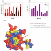
(A) Comparative analysis of the average SASA for each residue in assembled and unassembled state. Black represents the assembled state while red represents the unassembled state. (B) ΔSASA (ΔSASA=SASAunassembled - SASAassembled) values for each residue. (C) Surface view of cluster illustrating polarities of residues with color discrimination.
Figure 8
Figure 8
(A) ThT fluorescence assay of hIAPP (black) solution incubated with 1 (red) and 2 (blue) equivalents of TK9; ThT aggregation of TK9 peptide in solution is shown in the green trace. (B) CD spectra of freshly dissolved hIAPP (black) and hIAPP incubated over 6 hours with 2 equivalents of TK9 at the indicated time intervals of aggregation. (C) One dimensional 1H NMR spectrum of freshly dissolved TK9 in hIAPP (monomer) (top, control); STD spectrum in the presence of hIAPP monomer at t = 12 h. (middle) and in the presence of hIAPP fibril (bottom).
Scheme 1
Scheme 1
(A) SARS CoV-E sequence with TK9 region shown in blue. (B) The three-dimensional solution structure of SARS CoV-E protein in sodium dodecyl sulfate (SDS) micelle (2MM4.pdb). The C-terminal tail, Thr55 – Lys 63 adopted alpha helical conformation in micelle, makerd by circle, used in this study. (C) The primary sequence of C-terminal tail, TK9 or its shorter fragments.

Similar articles

Cited by

References

    1. Gazit E. Self-assembled peptide nanostructures: the design of molecular building blocks and their technological utilization. Chem. Soc. Rev. 2007;36:1263–1269. - PubMed
    1. Zhao X, Pan F, Xu H, Yaseen M, Shan H, Hauser CA, Zhang S, Lu JR. Molecular self-assembly and applications of designer peptide amphiphiles. Chem. Soc. Rev. 2010;39:3480–3498. - PubMed
    1. Zhang S. Fabrication of novel biomaterials through molecular self-assembly. Nat. Biotechnol. 2003;21:1171–1178. - PubMed
    1. Whitesides G, Mathias J, Seto C. Molecular self-assembly and nanochemistry: a chemical strategy for the synthesis of nanostructures. Science. 1991;254:1312–1319. - PubMed
    1. Lecommandoux S, Achard M-F, Langenwalter JF, Klok H-A. Self-assembly of rod-coil diblock oligomers based on α-helical peptides. Macromolecules. 2001;34:9100–9111.

Publication types

MeSH terms