Review
. 2015 Mar 17;42(3):406-17.
doi: 10.1016/j.immuni.2015.02.002.

Mitochondria in the regulation of innate and adaptive immunity

Affiliations
Review

Mitochondria in the regulation of innate and adaptive immunity

Samuel E Weinberg et al. Immunity. .

Abstract

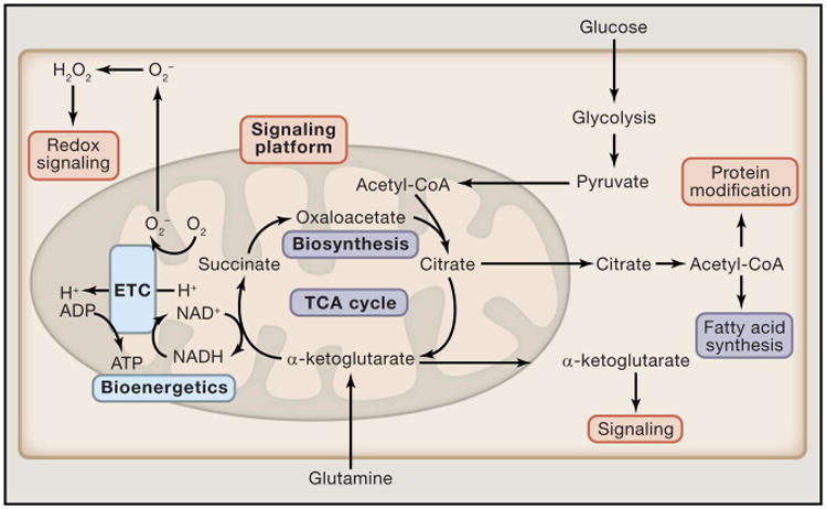
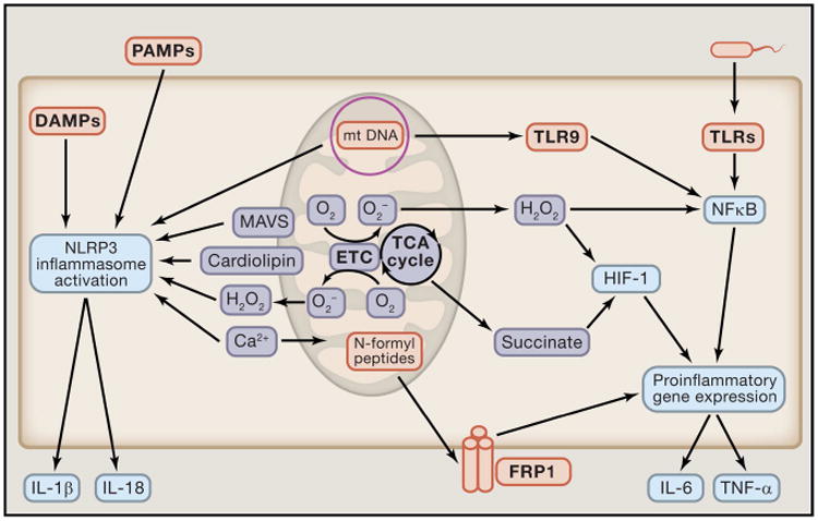
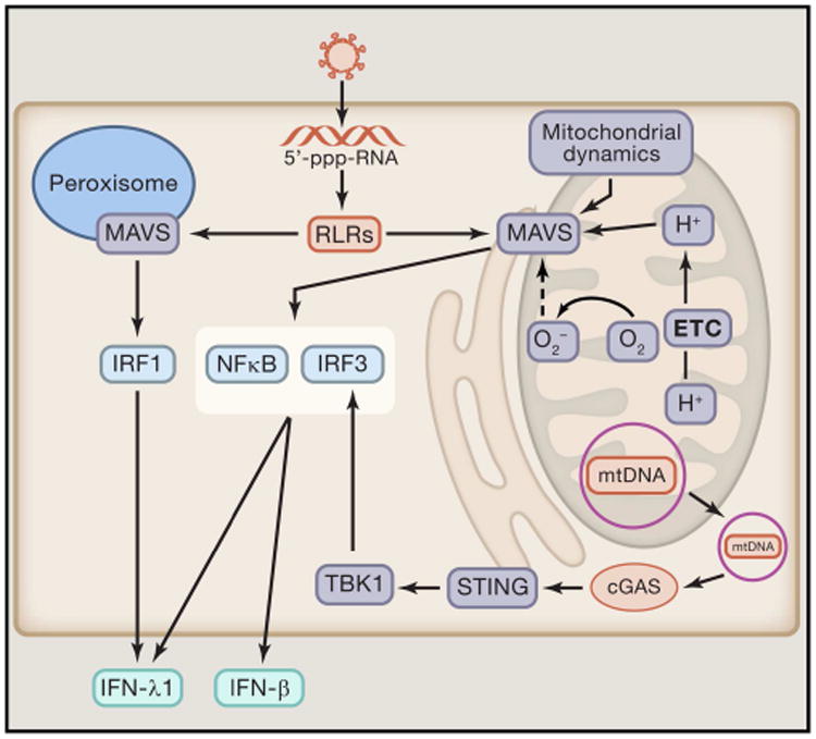
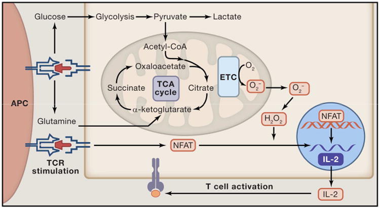
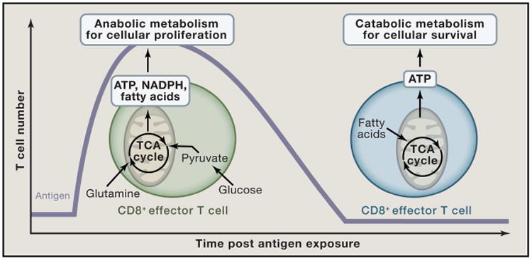
Mitochondria are well appreciated for their role as biosynthetic and bioenergetic organelles. In the past two decades, mitochondria have emerged as signaling organelles that contribute critical decisions about cell proliferation, death, and differentiation. Mitochondria not only sustain immune cell phenotypes but also are necessary for establishing immune cell phenotype and their function. Mitochondria can rapidly switch from primarily being catabolic organelles generating ATP to anabolic organelles that generate both ATP and building blocks for macromolecule synthesis. This enables them to fulfill appropriate metabolic demands of different immune cells. Mitochondria have multiple mechanisms that allow them to activate signaling pathways in the cytosol including altering in AMP/ATP ratio, the release of ROS and TCA cycle metabolites, as well as the localization of immune regulatory proteins on the outer mitochondrial membrane. In this Review, we discuss the evidence and mechanisms that mitochondrial dependent signaling controls innate and adaptive immune responses.


Figures

Figure 1
Figure 1. Mitochondria are essential metabolic and signaling organelles
Cytosolic metabolic pathways funnel into the mitochondria where they constantly replenish the TCA cycle. Depending on the cellular metabolic state, TCA cycle intermediates can be further oxidized to generate ATP (Blue) or they can be shuttled out of the mitochondria into subsidiary pathways to generate cellular building blocks such as fatty acids (purple). Finally, TCA cycle metabolites (Orange) in addition to other byproducts of mitochondrial metabolism, such as ROS, function as important signaling molecules which control cellular functions.
Figure 2
Figure 2. Mitochondria are critical to activation of the immune response
Mitochondrial components such as mitochondrial DNA (mtDNA) and N-formyl peptides can act as damage-associated molecular patterns (DAMPs). Specifically, mtDNA can activate the NLRP3 inflammasome and toll-like receptor-9 (TLR-9) to induce an inflammatory response, while N-formyl peptides activate pro-inflammatory gene expression through the N-formyl peptide receptor-1 (FRP1). In addition to production and presentation of DAMPs, mitochondrial metabolism and signaling further promote the induction of inflammation to pathogens. Mitochondrial derived metabolites such as mROS and succinate enhance pro-inflammatory gene expression, and mROS also can function directly as an anti-microbial effector molecule and NLRP3 activator. Additionally, mitochondrial localized proteins and lipids, MAVS and cardiolipin respectively, are required for proper activation of the NRLP3 inflammasome. Finally, cellular calcium flux is linked to NLRP3 activation and mitochondrial function and signaling.
Figure 3
Figure 3. The mitochondria are essential for the proper induction of antiviral signaling
The mitochondrial antiviral signaling protein (MAVS) is required for proper induction of RLR mediated activation of antiviral immunity. Importantly, mitochondrial dynamics, membrane potential, and ROS production are critical regulators of MAVS signaling. MAVS also localizes to the peroxisome, an organelle with a well described role in fatty acid oxidation and H2O2 generation. Stimulation of peroxisomal MAVS signaling induces IFN-λ1 production through IRF1 activation, while mitochondrial localized MAVS drives IFN-β and IFN-λ1 production. Along with MAVS, the mitochondrial also function as an activator of the stimulator of interferon genes protein (STING). Specifically, mtDNA translocated to the cytoplasm activates cyclic GMP-AMP synthase (cGAS) which triggers STING signaling which further enhances antiviral
Figure 4
Figure 4. Mitochondrial signaling is required for T cell activation
Upon binding of the TCR with MHC, numerous signaling cascades are activated. One of the activated pathways MYC activates an anabolic metabolic program that increases uptake of glucose and glutamine and allows for the cells to meet the increased metabolic demands of proliferation and induction of an adaptive immune response (Blue). Importantly, in this model metabolism works to sustain cellular activity required for a proper immune response. However, the mitochondria also alter cellular signaling upon ligation of the TCR. Specifically, mitochondrial ROS production following TCR stimulation is required for proper activation of NFAT and IL-2 production (Orange). In this way, the mitochondrial signaling and metabolism are required for proper T cell activation.
Figure 5
Figure 5. CD8+ effector T cells display a distinct mitochondrial metabolic profile compared to CD8+ memory T cells
Upon T cell activation, effector T cells rapidly increase uptake of glucose and glutamine which is utilized to produce the ATP and cellular building blocks (NADPH and fatty acids) required for production of clonal T cells. The mitochondria in effector T cells functions as an anabolic hub where TCA cycle intermediates are shuttled into the cytoplasm to promote production of increase cellular biomass. In contrast, memory T cells utilize fatty acid catabolism to efficiently generate ATP to fuel cellular survival.

References

    1. Barber GN. STING-dependent cytosolic DNA sensing pathways. Trends Immunol. 2014;35:88–93. - PubMed
    1. Berod L, Friedrich C, Nandan A, Freitag J, Hagemann S, Harmrolfs K, Sandouk A, Hesse C, Castro CN, Bähre H, et al. De novo fatty acid synthesis controls the fate between regulatory T and T helper 17 cells. Nat Med. 2014;20:1327–1333. - PubMed
    1. Bulua AC, Simon A, Maddipati R, Pelletier M, Park H, Kim KY, Sack MN, Kastner DL, Siegel RM. Mitochondrial reactive oxygen species promote production of proinflammatory cytokines and are elevated in TNFR1-associated periodic syndrome (TRAPS) J Exp Med. 2011;208:519–533. - PMC - PubMed
    1. Camões F, Bonekamp NA, Delille HK, Schrader M. Organelle dynamics and dysfunction: A closer link between peroxisomes and mitochondria. J Inherit Metab Dis. 2009;32:163–180. - PubMed
    1. Capasso M, Bhamrah MK, Henley T, Boyd RS, Langlais C, Cain K, Dinsdale D, Pulford K, Khan M, Musset B, et al. HVCN1 modulates BCR signal strength via regulation of BCR-dependent generation of reactive oxygen species. Nat Immunol. 2010;11:265–272. - PMC - PubMed

Publication types

MeSH terms