. 2015 Jan 30;21(1):109-22.
doi: 10.2119/molmed.2014.00158.

Investigation on the Origin of Sperm DNA Fragmentation: Role of Apoptosis, Immaturity and Oxidative Stress

Affiliations

Investigation on the Origin of Sperm DNA Fragmentation: Role of Apoptosis, Immaturity and Oxidative Stress

Monica Muratori et al. Mol Med. .

Abstract

Sperm DNA fragmentation (sDF) represents a threat to male fertility, human reproduction and the health of the offspring. The causes of sDF are still unclear, even if apoptosis, oxidative assault and defects in chromatin maturation are hypothesized. Using multicolor flow cytometry and sperm sorting, we challenged the three hypothesized mechanisms by simultaneously evaluating sDF and signs of oxidative damage (8-hydroxy, 2'-deoxyguanosine [8-OHdG] and malondialdehyde [MDA]), apoptosis (caspase activity and cleaved poly[ADP-ribose] polymerase [cPARP]) and sperm immaturity (creatine phosphokinase [CK] and excess of residual histones). Active caspases and c-PARP were concomitant with sDF in a high percentage of spermatozoa (82.6% ± 9.1% and 53.5% ± 16.4%, respectively). Excess of residual histones was significantly higher in DNA-fragmented sperm versus sperm without DNA fragmentation (74.8% ± 17.5% and 37.3% ± 16.6%, respectively, p < 0.005), and largely concomitant with active caspases. Conversely, oxidative damage was scarcely concomitant with sDF in the total sperm population, at variance with live sperm, where 8-OHdG and MDA were clearly associated to sDF. In addition, most live cells with active caspase also showed 8-OHdG, suggesting activation of apoptotic pathways in oxidative-injured live cells. This is the first investigation on the origin of sDF directly evaluating the simultaneous presence of the signs of the hypothesized mechanisms with DNA breaks at the single cell level. The results indicate that the main pathway leading to sperm DNA breaks is a process of apoptosis, likely triggered by an impairment of chromatin maturation in the testis and by oxidative stress during the transit in the male genital tract. These findings are highly relevant for clinical studies on the effects of drugs on sDF and oxidative stress in infertile men and for the development of new therapeutic strategies.


Figures

Figure 1
Figure 1
(A) Typical FSC/SSC dot plot of a semen sample showing the flame-shaped region (FR) containing semen apoptotic bodies and sperm. The latter are defined by nuclear staining with DAPI (B), that also distinguishes two sperm populations named brighter and dimmer because of the different intensity of nuclear staining. Total sperm population is included in R1 region, whereas brighter sperm are in R2 region.
Figure 2
Figure 2
Concomitance of sDF and CK in human spermatozoa. (A) Dot plots of the negative control (left panel) and the corresponding test sample (right panel) showing the percentages of sperm without sDF and CK (LL quadrant), sperm with sDF and without CK (UL quadrant), sperm with sDF and CK (UR quadrant) and sperm without sDF and with CK (LR quadrant). Representative of nine experiments. Percentages in the quadrants refer to the shown sample. (B) Percentage of CK in fragmented (UR/[UR + UL]) and nonfragmented (LR/[LL + LR]) sperm. (C) Images from fluorescence microscopy showing the localization of CK (left panel) in sperm with and without TUNEL labeling (middle panel). Right panel: bright field. Original magnification 1,000×. LL, lower left; UL, upper left; LR, lower right; UR, upper right.
Figure 3
Figure 3
Sorting fragmented and nonfragmented sperm by FACSAria II and staining with AB (n = 5). (A) TUNEL fluorescence histograms of the sorted fragmented and nonfragmented sperm. (B) Staining with AB of the sorted fractions. Sperm nuclei with intense dark-blue staining were considered to be immature. Original magnification 1,000×. (C) Percentage of chromatin immaturity, as detected by AB staining, in fragmented and nonfragmented sperm. **p < 0.01.
Figure 4
Figure 4
Concomitance of sDF and active caspases in human spermatozoa. (A) Dot plots of the negative control (left panel) and the corresponding test sample (right panel) showing the percentages of sperm without sDF and active caspases (LL quadrant), sperm with sDF and without active caspases (UL quadrant), sperm with sDF and caspase activity (UR quadrant) and sperm without sDF and with active caspases (LR quadrant). Representative of 13 experiments. Percentages in the quadrants refer to the shown sample. (B) Percentage of sperm with active caspases in fragmented (UR/[UR + UL]) and nonfragmented (LR/[LL + LR]) sperm. (C) Images from fluorescence microscopy showing the localization of active caspases (left panel) in sperm with and without TUNEL labeling (middle panel). Right panel: bright field. Original magnification 1,000×. ***p < 0.001. LL, lower left; UL, upper left; LR, lower right; UR, upper right.
Figure 5
Figure 5
Concomitance of sDF and cPARP in human spermatozoa. (A) Dot plots of the negative control (left panel) and the corresponding test sample (right panel) showing the percentages of sperm without sDF and cPARP (LL quadrant), sperm with sDF and without cPARP (UL quadrant), sperm with sDF and cPARP (UR quadrant), sperm without sDF and with cPARP (LR quadrant). Representative of 10 experiments. Percentages in the quadrants refer to the shown sample. (B) Percentage of sperm with cPARP in fragmented (UR/[UR + UL]) and nonfragmented (LR/[LL + LR]) sperm. (C) Images from fluorescence microscopy showing the localization of cPARP (left panel) in sperm with and without TUNEL labeling (middle panel). Right panel: bright field. Original magnification 1,000×. ***p < 0.001. LL, lower left; UL, upper left; LR, lower right; UR, upper right.
Figure 6
Figure 6
Concomitance of sDF and oxidative signs in human spermatozoa. 8-OHdG: (A) Dot plots of the negative control (left panel) and the corresponding test sample (right panel) showing the percentages of sperm without sDF and 8-OHdG (LL quadrant), sperm with sDF and without 8-OHdG (UL quadrant), sperm with sDF and 8-OHdG (UR quadrant) and sperm without sDF and with 8-OHdG (LR quadrant). Representative of 21 experiments. Percentages in the quadrants refer to the shown sample. (B) Percentage of sperm with 8-OHdG in fragmented (UR/[UR + UL]) and nonfragmented (LR/[LL + LR]) sperm, in total (left) and brighter (right) sperm population. MDA: (C) Dot plots of the negative control (left panel) and the corresponding test sample (right panel) showing the percentages of sperm without sDF and MDA (LL quadrant), sperm with sDF and without MDA (UL quadrant), sperm with sDF and MDA (UR quadrant) and sperm without sDF and with MDA (LR quadrant). Representative of 11 experiments. Percentages in the quadrants refer to the shown sample. (D) Percentage of sperm with MDA in fragmented sperm (UR/[UR + UL]) and nonfragmented (LR/[LL + LR]) sperm, in total (left) and brighter (right) sperm population. (E) Images from fluorescence microscopy showing the localization of MDA (left panel) in sperm with and without TUNEL labeling (middle panel). Right panel: bright field. Original magnification 1,000×. *p < 0.05; ***p < 0.001. LL, lower left; UL, upper left; LR, lower right; UR, upper right.
Figure 7
Figure 7
Experiments in live sperm. (A) Concomitance of sDF and 8-OHdG (n = 6). Dot plots of the negative control (upper panel) and the corresponding test sample (middle panel) showing percentage of: sperm without sDF and 8-OHdG (LL quadrant), sperm with sDF and without 8-OHdG (UL quadrant), sperm with sDF and 8-OHdG (UR quadrant), sperm without sDF and with 8-OHdG (LR quadrant). Representative of six experiments. Percentages in the quadrants refer to the shown sample. Lower panel, percentage of live sperm with 8-OHdG in fragmented (UR/[UR + UL]) and nonfragmented sperm (LR/[LL + LR]). (B) Concomitance of sDF and MDA (n = 6). Dot plots of the negative control (upper panel) and the corresponding test sample (middle panel) showing sperm without sDF and MDA (LL quadrant), sperm with sDF and without MDA (UL quadrant), sperm with sDF and MDA (UR quadrant) and sperm without sDF and with MDA (LR quadrant). Representative of six experiments. Percentages in the quadrants refer to the shown sample. Lower panel, percentage of live sperm with MDA in fragmented (UR/[UR + UL]) and nonfragmented sperm (LR/[LL + LR]). (C) Concomitance of sDF and active caspases (n = 7). Dot plots of the negative control (upper panel) and the corresponding test sample (middle panel) showing sperm without sDF and active caspases (LL quadrant), sperm with sDF and without active caspases (UL quadrant), sperm with sDF and active caspases (UR quadrant) and sperm without sDF and with active caspases (LR quadrant). Representative of six experiments. Percentages in the quadrants are referred to the shown sample. Lower panel, percentage of live sperm with active caspases in fragmented (UR/[UR + UL]) and nonfragmented sperm (LR/[LL + LR]). (D) Concomitance of 8-OHdG and active caspases (n = 3). Dot plots of the negative control (left panel) and the corresponding test sample (right panel) showing sperm without 8-OHdG and active caspases (LL quadrant), sperm with 8-OHdG and without active caspases (UL quadrant), sperm with 8-OHdG and active caspases (UR quadrant) and sperm without 8-OHdG and with active caspases (LR quadrant). Representative of three experiments. Percentages in the quadrants refer to the shown sample. *p < 0.05; **p < 0.01. LL, lower left; UL, upper left; LR, lower right; UR, upper right.
Figure 8
Figure 8
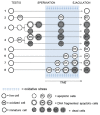
A scheme to summarize the results of the study. Lane 1: Live cells arriving in the ejaculate without DNA damage. Lane 2: Immature live cells arriving in the ejaculate without DNA damage. Lanes 3 and 4: Apoptotic cells evolving as DNA fragmented and dead (or directly dead). In Lane 4, apoptosis is induced by maturation impairment. These cells go through the genital tracts without being further DNA damaged by oxidative stress. Lane 5: Live cells undergoing oxidative damage just before ejaculation and thus not experiencing apoptosis. Lane 6: Live cells undergoing oxidative damage triggering apoptosis but not DNA fragmentation. Lane 7: Live cells undergoing oxidative damage triggering apoptosis and DNA fragmentation. Lane 8: Live cells undergoing oxidative damage triggering apoptosis, DNA fragmentation and death.

References

    1. Ahmadi A, Ng SC. Developmental capacity of damaged spermatozoa. Hum Reprod. 1999;14:2279–85. - PubMed
    1. Singh NP, et al. Abundant alkali-sensitive sites in DNA of human and mouse sperm. Exp Cell Res. 1989;184:461–70. - PubMed
    1. Gorczyca W, Traganos F, Jesionowska H, Darzynkiewicz Z. Presence of DNA strand breaks and increased sensitivity of DNA in situ to denaturation in abnormal human sperm cells: analogy to apoptosis of somatic cells. Exp Cell Res. 1993;207:202–5. - PubMed
    1. Tamburrino L, et al. Mechanisms and clinical correlates of sperm DNA damage. Asian J Androl. 2012;14:24–31. - PMC - PubMed
    1. Morris ID. Sperm DNA damage and cancer treatment. Int J Androl. 2002;25:255–61. - PubMed

Publication types