. 2015 May;200(1):207-19.
doi: 10.1534/genetics.115.176180. Epub 2015 Mar 18.

The aggregation-prone intracellular serpin SRP-2 fails to transit the ER in Caenorhabditis elegans

Affiliations

The aggregation-prone intracellular serpin SRP-2 fails to transit the ER in Caenorhabditis elegans

Richard M Silverman et al. Genetics. 2015 May.

Abstract

Familial encephalopathy with neuroserpin inclusions bodies (FENIB) is a serpinopathy that induces a rare form of presenile dementia. Neuroserpin contains a classical signal peptide and like all extracellular serine proteinase inhibitors (serpins) is secreted via the endoplasmic reticulum (ER)-Golgi pathway. The disease phenotype is due to gain-of-function missense mutations that cause neuroserpin to misfold and aggregate within the ER. In a previous study, nematodes expressing a homologous mutation in the endogenous Caenorhabditis elegans serpin, srp-2, were reported to model the ER proteotoxicity induced by an allele of mutant neuroserpin. Our results suggest that SRP-2 lacks a classical N-terminal signal peptide and is a member of the intracellular serpin family. Using confocal imaging and an ER colocalization marker, we confirmed that GFP-tagged wild-type SRP-2 localized to the cytosol and not the ER. Similarly, the aggregation-prone SRP-2 mutant formed intracellular inclusions that localized to the cytosol. Interestingly, wild-type SRP-2, targeted to the ER by fusion to a cleavable N-terminal signal peptide, failed to be secreted and accumulated within the ER lumen. This ER retention phenotype is typical of other obligate intracellular serpins forced to translocate across the ER membrane. Neuroserpin is a secreted protein that inhibits trypsin-like proteinase. SRP-2 is a cytosolic serpin that inhibits lysosomal cysteine peptidases. We concluded that SRP-2 is neither an ortholog nor a functional homolog of neuroserpin. Furthermore, animals expressing an aggregation-prone mutation in SRP-2 do not model the ER proteotoxicity associated with FENIB.

Keywords: ER stress; SRP-2; familial encephalopathy with neuroserpin inclusions; neuroserpin; proteostasis.


Figures

Figure 1
Figure 1
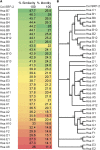
C. elegans SRP-2 protein sequence compared to human serpin family members. (A) Percentage of similarity and identity scores of the primary amino acid sequence of SRP-2 compared to the 36 human (Hsa) members. Human serpins are listed in order of increasing to decreasing percentage of similarity and have the prefix SERPIN before the clade and member number omitted. (B) An unrooted, uncorrected phylogram of the multiple-sequence alignment performed using the neighbor-joining method and bootstrapped 1000 times. Numbers at branch points indicate the percentage that those branch points occurred in the 1000 generated trees.
Figure 2
Figure 2
Confocal image analysis of transgenic animals expressing wild-type and mutant (± s)SRP-2::GFP transgenes. Shown are GFP and DsRed fluorescence images of animals coexpressing sDsRed::KDEL and sGFP::ATZ (A–D), GFP::ATZ (F–I), SRP-2::GFP (K–N), sSRP-2::GFP (P–S), SRP-2H302R::GFP (U–X), and sSRP-2H302R::GFP (Z–CC) under the control of the intestinal-specific promoter from nhx-2. White arrowheads highlight regions that colocalize in the GFP (A, P, and Z) and DsRed (B, Q, and AA) channels. White arrows highlight regions that do not colocalize in the GFP (U) and DsRed (W) channels. Regions highlighted by white boxes are shown at higher magnifications as merged images (D, I, N, S, X, and CC). Colocalization plots were generated using the colocalization feature in the Volocity package. Images were likely colocalized if the colocalization plot showed a linear relationship and the Pearson’s correlation coefficient was >0.750 (Barlow et al. 2010).
Figure 3
Figure 3
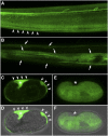
Comparison of confocal images from transgenic animals expressing a classical secreted serpin, sGFP::ATM vs. the intracellular serpin, SRP-2::GFP. (A and B) Maximum-intensity projection fluorescence images of the posterior region of animals expressing sGFP::ATM (A) and SRP-2::GFP (B) under the control of the srp-2 promoter. The srp-2 promoter is primarily active in hypodermal cells throughout development (Pak et al. 2004). The hypodermis is responsible for synthesizing the cuticle and secreted products become incorporated or trapped within this structure. As shown previously (Long et al. 2014) and reimaged here for comparison, sGFP::ATM expressed under the control of the srp-2 promoter showed little hypodermal retention and instead accumulated within the cuticle of the worm (A, arrowheads). These results suggested that sGFP::ATM was efficiently secreted by the hypodermal cells. Unlike sGFP::ATM, however, SRP-2::GFP was detected within hypodermal cells (B, arrows), but no protein was detected beneath or within the cuticle. These results further support our findings suggesting that SRP-2 is an obligate intracellular serpin. We also examined comma-stage embryos from transgenic animals expressing sGFP::ATM (C and D) (DIC merge) or SRP-2::GFP (E and F) (DIC merge). sGFP::ATM was rapidly secreted and mostly visible within the extracellular perivitelline space between the embryo and the egg shell (C and D, asterisk and arrowheads). In contrast, SRP-2::GFP was visible only within cells. These images confirmed that wild-type SRP-2 localized to the cytosol and there was no detectable secretion.

References

    1. Amen T., Kaganovich D., 2014. Dynamic droplets: the role of cytoplasmic inclusions in stress, function, and disease. Cell. Mol. Life Sci. 72: 401–415. - PMC - PubMed
    1. Barlow A. L., Macleod A., Noppen S., Sanderson J., Guerin C. J., 2010. Colocalization analysis in fluorescence micrographs: verification of a more accurate calculation of Pearson’s correlation coefficient. Microsc. Microanal. 16: 710–724. - PubMed
    1. Belin D., 1993. Biology and facultative secretion of plasminogen activator inhibitor-2. Thromb. Haemost. 70: 144–147. - PubMed
    1. Belin D., Guzman L. M., Bost S., Konakova M., Silva F., et al. , 2004. Functional activity of eukaryotic signal sequences in Escherichia coli: the ovalbumin family of serine protease inhibitors. J. Mol. Biol. 335: 437–453. - PubMed
    1. Benyair R., Ogen-Shtern N., Lederkremer G. Z., 2014. Glycan regulation of ER-associated degradation through compartmentalization. Semin. Cell Dev. Biol. (in press). - PubMed

Publication types

MeSH terms

LinkOut - more resources