Review
. 2015 Jul;104(4):213-46.
doi: 10.1002/bip.22643.

KDM1 class flavin-dependent protein lysine demethylases

Affiliations
Review

KDM1 class flavin-dependent protein lysine demethylases

Jonathan M Burg et al. Biopolymers. 2015 Jul.

Abstract

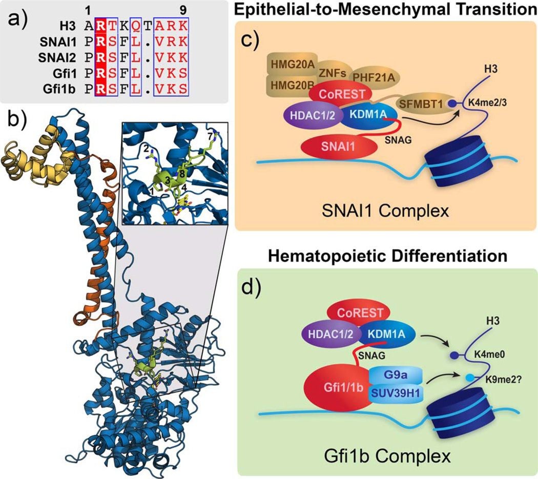
Flavin-dependent, lysine-specific protein demethylases (KDM1s) are a subfamily of amine oxidases that catalyze the selective posttranslational oxidative demethylation of methyllysine side chains within protein and peptide substrates. KDM1s participate in the widespread epigenetic regulation of both normal and disease state transcriptional programs. Their activities are central to various cellular functions, such as hematopoietic and neuronal differentiation, cancer proliferation and metastasis, and viral lytic replication and establishment of latency. Interestingly, KDM1s function as catalytic subunits within complexes with coregulatory molecules that modulate enzymatic activity of the demethylases and coordinate their access to specific substrates at distinct sites within the cell and chromatin. Although several classes of KDM1-selective small molecule inhibitors have been recently developed, these pan-active site inhibition strategies lack the ability to selectively discriminate between KDM1 activity in specific, and occasionally opposing, functional contexts within these complexes. Here we review the discovery of this class of demethylases, their structures, chemical mechanisms, and specificity. Additionally, we review inhibition of this class of enzymes as well as emerging interactions with coregulatory molecules that regulate demethylase activity in highly specific functional contexts of biological and potential therapeutic importance.

Keywords: Epigenetic; KDM1A; KDM1B; LSD1; LSD2.


Figures

FIGURE 1
FIGURE 1
Structural overview of KDM1 demethylases. (a) Sequence alignment of a portion of the amine oxidase domains of KDM1A, KDM1B, MAO-A, MAO-B, and maize polyamine oxidase (mPAO). Sequence conserved active site Lys residue is starred. (b) Domain maps of KDM1A and KDM1B. SWI3p, Rsc8p, and Moira (SWIRM) domains shown in green, amine oxidase domains shown in blue, tower domain shown in lavender, linker domain shown in teal, C4H2C2 domain shown in purple, and Zf-CW domain shown in orange. (c) Ribbon representation of the structure of KDM1A. Domains follow same color scheme as outlined above (PDB 2IW5). (d) Ribbon representation of the structure of KDM1B. Domains follow same color scheme as outlined above (PDB 4HSU). (e) Venn diagram of domain conservation between KDM1A and KDM1B.
FIGURE 2
FIGURE 2
Overview of structural conformations of bound peptide ligands. (a) Conformation of covalently bound N-methypropargyl-Lys4 H3 peptide in KDM1A (PDB 2UXN). (b) Conformation of non-covalently bound H3K4M (pK4M) in KDM1A (PDB 2V1D). (c) Conformation of non-covalently bound SNAI1 in KDM1A (PDB 2Y48). (d) Conformation of non-covalently bound pK4M in KDM1B (PDB 4HSU). (e) Structural alignment of pK4M and SNAI1 (b and c, respectively) bound to KDM1A, illustrating the overall fold conservation of the peptides and relative position of the non-covalent FAD and active-site Lys661.
FIGURE 3
FIGURE 3
Chemical structures of representative mechanism-based, irreversible inhibitors.
FIGURE 4
FIGURE 4
Structures of substrate-based peptide inhibitors.
FIGURE 5
FIGURE 5
Structures of nonpeptidic, warhead small molecule inhibitors that are substrate derived mimics.
FIGURE 6
FIGURE 6
Structures of bisguanide and bisguanidine polyamine inhibitors.
FIGURE 7
FIGURE 7
Structures of bifunctional small molecule inhibitors that incorporate multiple pharmacaphores.
FIGURE 8
FIGURE 8
Natural polyphenol inhibitors.
FIGURE 9
FIGURE 9
Structures of screen identified and validated inhibitors.
FIGURE 10
FIGURE 10
Structures of mechanism-based phenelzine analogs.
FIGURE 11
FIGURE 11
KDM1A complexes that utilize CoREST and CoREST-like interactions. (a) Ribbon representation of KDM1A (blue) in complex with the linker (orange) and SANT2 (yellow) domains of CoREST (PDB 2IW5). Insert highlights the regions of contact between CoREST and the KDM1A tower domain. (b) CoREST recruits a KDM1A complex to suppress neuronal genes through its interaction with REST, which also recruits the Sin3 complex. (c) The CoREST core complex is recruited by a direct interaction between TLX and KDM1A. This complex forms a negative feedback loop with the microRNA miR-137. (d) The transcription factor TAL1 recruits the CoREST complex, and potentially other coregulators, to repress gene transcription as an effector of hematopoietic differentiation. (e) The CtBP proteins associate with DNA-binding proteins for genomic localization and recruit chromatin remodelers, including KDM1A, to regulate the EMT. (f) Domain maps of CoREST and MTA1 are representative of their respective isoforms and show a similar ELM2/SANT domain organization. g) KDM1A is recruited by its tower domain to the MTA subunit of the NuRD complex, and in this context suppresses the EMT in breast cancer.
FIGURE 12
FIGURE 12
KDM1A involvement in HSV infection and latency. KDM1A, along with other host cell epigenetic machinery, is coopted by HSV for viral gene activation. KDM1A and the CoREST/ REST/HDAC complex may also participate in viral gene repression, where KDM1A may demethylate H3K4 residues.
FIGURE 13
FIGURE 13
KDM1A as an effector of gene activation and repression with nuclear hormone receptors. (a) KDM1A promotes AR gene activation following hormone stimulation at androgen-responsive gene promoters. At high hormone levels, KDM1A, and potentially other corepressors, repress AR transcription by demethylating H3K4 residues in the corresponding enhancer region. (b) KDM1A and PELP1 are recruited to ERα-occupied promoters. PELP1 reads H3K4me2 marks, and may position KDM1A for H3K9 demethylation. Association of CAC1 with this ERα/KDM1A complex causes dissociation from the promoter, accumulation of H3K9me2 marks, and gene repression.
FIGURE 14
FIGURE 14
SNAG family proteins recruit KDM1A using product mimicry. (a) Alignment of the H3 tail with the first 8 residues of the SNAI1/2 and Gfi1/1b SNAG domains shows striking similarity, with conserved residues highlighted in red and chemically similar residues boxed in blue. (b) Ribbon representation of the ternary complex formed by KDM1A (blue), CoREST (orange/yellow), and SNAG (green; PDB 2Y48). The insert shows an enlarged view of the SNAG peptide in the KDM1A active site with numbered residues starting with the N-terminal proline. (c) SNAI1 recruits the CoREST core complex and other proteins through its SNAG domain to regulate the EMT. SFMBT1 may also help localize the complex by binding H3K4me2/3. (d) Gfi1 and Gfi1b also recruits the CoREST core complex and cooperates with HMTs to regulate hematopoietic processes.
FIGURE 15
FIGURE 15
KDM1A forms multi-component complexes with lncRNAs. (a) The lncRNA HOTAIR colocalizes the RE1-silencing transcription factor (REST)/CoREST/KDM1A and PRC2 complexes within the genome for subsequent gene repression. (b) While TRF2 protects telomere ends from degradation, loss of TRF2 allows recruitment of KDM1A and the telomeric repeat-containing lncRNA TERRA to the nuclease MRE11 and promotes telomere end processing.
FIGURE 16
FIGURE 16
KDM1B forms a multiprotein complex that epigenetically regulates gene expression by altering histone modifications in the coding regions of genes. (a) KDM1B associates with the chromatin reader Nuclear Protein 60kDa/Glyoxylate Reductase 1 Homolog (NPAC/GLYR1), RNA polymerase II, and other elongation factors within active genes. (b) A ribbon representation of KDM1B (colored as described in Figure 1) in complex with a NPAC/GLYR1 fragment peptide (pink). The insert shows an enlarge view of NPAC binding in the cleft between the amine oxidase catalytic domain and SWIRM domains of KDM1B.
SCHEME 1
SCHEME 1
Proposed chemical mechanism of FAD-dependent demethylases KDM1A and KDM1B. Oxidation of the C-N bond of the methylated lysine sidechain to an iminium ion, with concurrent reduction of the flavin enables hydrolysis via bulk water. Collapse of the hemiaminal (not shown) yields formaldehyde and the demethylated amine sidechain. The reduced flavin is reoxidized by molecular oxygen, generating a molar equivalent of hydrogen peroxide.

References

    1. Allfrey VG, Mirsky AE. Science. 1964;144:559. - PubMed
    1. Allfrey VG, Faulkner R, Mirsky AE. Proc Natl Acad Sci USA. 1964;51:786–794. - PMC - PubMed
    1. Kooistra SM, Helin K. Nat Rev Mol Cell Biol. 2012;13:297–311. - PubMed
    1. Hou H, Yu H. Curr Opin Struct Biol. 2010;20:739–748. - PMC - PubMed
    1. Qian C, Zhou MM. Cell Mol Life Sci. 2006;63:2755–2763. - PMC - PubMed

Publication types

MeSH terms

LinkOut - more resources