. 2015 May;89(10):5222-37.
doi: 10.1128/JVI.03614-14. Epub 2015 Mar 18.

Genome diversity of Epstein-Barr virus from multiple tumor types and normal infection

Affiliations

Genome diversity of Epstein-Barr virus from multiple tumor types and normal infection

Anne L Palser et al. J Virol. 2015 May.

Abstract

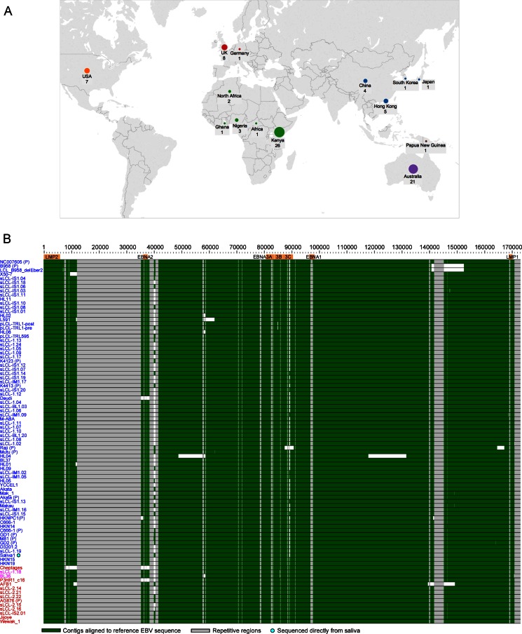
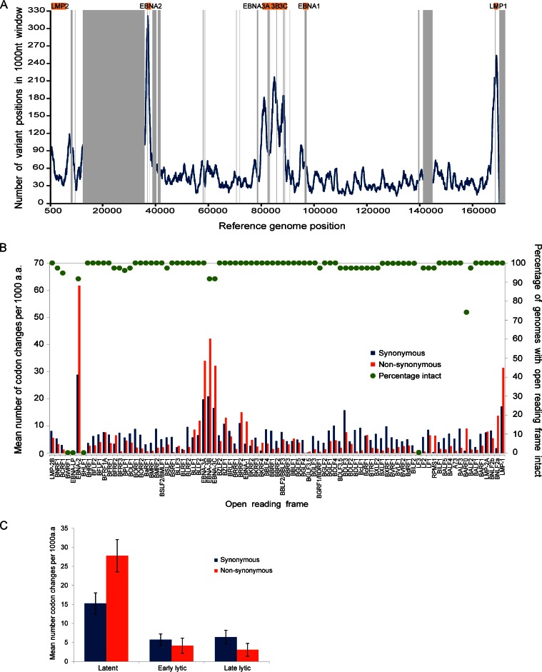
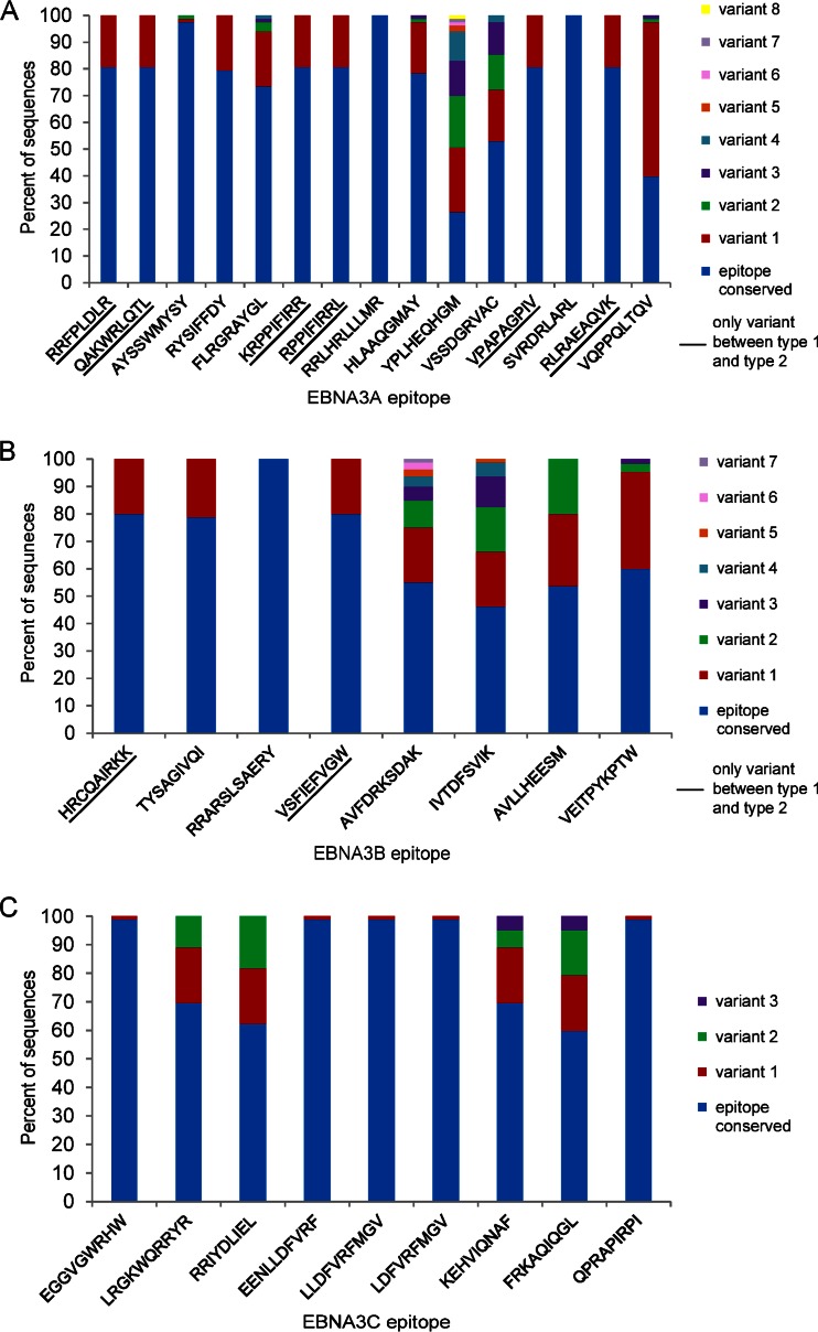
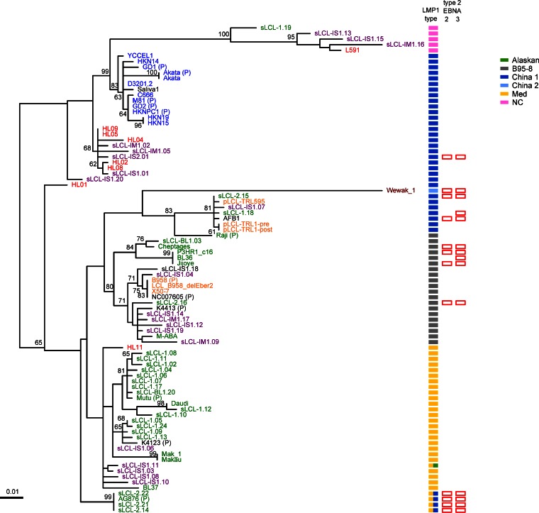
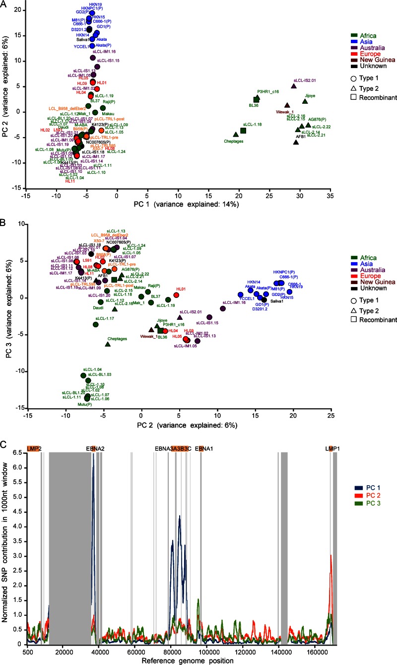
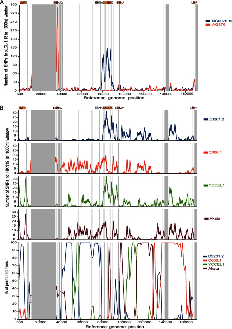
Epstein-Barr virus (EBV) infects most of the world's population and is causally associated with several human cancers, but little is known about how EBV genetic variation might influence infection or EBV-associated disease. There are currently no published wild-type EBV genome sequences from a healthy individual and very few genomes from EBV-associated diseases. We have sequenced 71 geographically distinct EBV strains from cell lines, multiple types of primary tumor, and blood samples and the first EBV genome from the saliva of a healthy carrier. We show that the established genome map of EBV accurately represents all strains sequenced, but novel deletions are present in a few isolates. We have increased the number of type 2 EBV genomes sequenced from one to 12 and establish that the type 1/type 2 classification is a major feature of EBV genome variation, defined almost exclusively by variation of EBNA2 and EBNA3 genes, but geographic variation is also present. Single nucleotide polymorphism (SNP) density varies substantially across all known open reading frames and is highest in latency-associated genes. Some T-cell epitope sequences in EBNA3 genes show extensive variation across strains, and we identify codons under positive selection, both important considerations for the development of vaccines and T-cell therapy. We also provide new evidence for recombination between strains, which provides a further mechanism for the generation of diversity. Our results provide the first global view of EBV sequence variation and demonstrate an effective method for sequencing large numbers of genomes to further understand the genetics of EBV infection.

Importance: Most people in the world are infected by Epstein-Barr virus (EBV), and it causes several human diseases, which occur at very different rates in different parts of the world and are linked to host immune system variation. Natural variation in EBV DNA sequence may be important for normal infection and for causing disease. Here we used rapid, cost-effective sequencing to determine 71 new EBV sequences from different sample types and locations worldwide. We showed geographic variation in EBV genomes and identified the most variable parts of the genome. We identified protein sequences that seem to have been selected by the host immune system and detected variability in known immune epitopes. This gives the first overview of EBV genome variation, important for designing vaccines and immune therapy for EBV, and provides techniques to investigate relationships between viral sequence variation and EBV-associated diseases.


Figures

FIG 1
FIG 1
Origin of samples and alignment of new EBV genome sequences. (A) World map depicting the origins of the 83 unique EBV genomes sequenced and analyzed in this study. The sizes of the circles are proportional to the number of samples from each geographic region, with country and number of genomes annotated. One sample without country information is listed as “African,” and one sample of unknown geographic origin is not shown. (B) Whole-genome multiple-sequence alignment of all EBV genomes, including the EBV (B95-8/Raji) reference genome (NC_007605, top) and 12 previously published genomes (annotated with a “P”). Strains are ordered by their similarity to the sequence of the type 1 reference strain (NC_007605), shown at the top. Type 1 genomes are in blue, type 2 genomes are in red, and recombinant (type 1/2) sequences are in purple. Contigs are indicated in green. Repetitive regions are masked out in gray. Gaps and deletions are in white. The wild-type genome derived from the saliva of a healthy individual is annotated with a turquoise dot. The HL04 Hodgkin lymphoma biopsy specimen has an inversion of a section of the genome from position 65000 to 125000. Known deletions in Daudi, P3HR1, pLCL-TRL1, and an LCL generated with a B95-8-based BAC (LCL delEBER2) were detected.
FIG 2
FIG 2
SNP variation across all EBV genomes. (A) Single nucleotide polymorphism (SNP) frequency across 83 unique EBV genomes. The line graph is plotted across the genome showing the number of base positions in a sliding 1,000-nt window where at least one EBV sequence has a SNP relative to the consensus sequence. Repeat regions are masked out in gray. (B) Mean number of codon changes (relative to the consensus of 83 EBV sequences) per gene across the genome, presented as codon changes per 1,000 amino acids to normalize for gene length. Synonymous nucleotide changes are indicated by blue bars and nonsynonymous changes by red bars. The repeat regions within BZLF1, BPLF1, BLLF1, and EBNA1, -2, -3B, and -3C have been masked, and data are provided for the nonrepetitive region only. The right scale shows the percentage of EBV genomes with an intact open reading frame for each gene. BWRF1, EBNA-LP, BHLF1, and LF3 were incompletely assembled due to repetitive regions and were not determined. (C) Numbers of codon changes per gene (means ± standard errors of the means, normalized per kilobase), separated into gene type. Latent genes have an increased number of changes compared to early and late lytic genes. Latency-associated genes also have an increased ratio of nonsynonymous to synonymous coding changes compared to lytic genes.
FIG 3
FIG 3
Variation in T-cell epitopes in EBNA3 genes. Variation in known T-cell epitopes in EBNA3A (A), EBNA3B (B) and EBNA3C (C) genes across all 83 EBV genomes. Graphs indicate the percentage of EBV genome sequences with the epitope fully conserved (blue) and the percentage of sequences that have each of the variant sequences as a stacked histogram. Some epitopes are fully conserved across all strains (fully blue bars), some have differences only between type 1 and type 2 (underlined), and some have multiple variants. Only sequences with an intact open reading frame were included.
FIG 4
FIG 4
LMP1 phylogenetic tree classified by LMP1 type. Maximum-likelihood phylogenetic tree of LMP1 nucleotide sequence from 83 EBV strains (71 new strains and 12 published strains, annotated with a “P”). Bootstrap values above 60 are shown, and the scale bar represents 0.01 nucleotide substitution per site. All strains could be classified into one of the 7 LMP1 types (as defined in reference ; see the key for colors), and these define the major clades of the tree, except China 1 strains, which are split across several clades. Strains with type 2 EBNA2 and EBNA3 (empty red bars) are present in multiple LMP1 types but absent from the NC and Med LMP1 types. Geographic origin (shown by the color of the sample name: blue, Asia; green, Africa; red, Europe; yellow, USA; purple, Australia; dark red, Papua New Guinea) shows that many of the LMP1 types, including Med and NC, are found in strains from a wide geographic area.
FIG 5
FIG 5
Principal-component analysis separates strains by type 1/2 and geographic origin. Principal-component analysis (PCA) of all EBV strains (71 new strains and 12 published strains, annotated with a “P”) based on SNPs relative to the consensus sequence in a full-genome multiple-sequence alignment. (A) Principal component 1 separates all strains based on type 1 and type 2, with all type 2 strains (triangles) clustering together (including two intertypic recombinants, BL36 and sLCL-1.18, which have type 2 EBNA3s). (B) Principal component 2 shows some geographic clustering of EBV strains, with clear separation of the Asian strains (blue). (C) Regions of the EBV genome that contribute to the first three principal components.
FIG 6
FIG 6
Recombination between different EBV strains. (A) Intertypic recombination between type 1 and type 2 EBV strains. Diversity plots show the number of SNPs per 1,000-nt sliding window comparing EBV strain sLCL-1.18 with the type 1 reference genome (NC_007605; blue) and the type 2 reference genome (AG876; red). Major repeat regions are masked out (vertical gray bars). The sLCL-1.18 EBV genome is highly similar to the type 1 genome in the EBNA2 region (around positions 36000 to 37000) and highly similar to type 2 in the EBNA3s region (around positions 80000 to 89000). (B) Evidence for recombination between different type 1 EBV strains. Diversity and bootscan plots comparing strain HKN19 with 4 geographically related (southeast Asian) EBV strains, with repeat regions masked out (gray bars). Diversity plots (top; number of SNPs in a 1,000-nt sliding window) show high levels of similarity (few SNPs) between HKN19 and strain D3201.2 until approximately position 80000 and similarity to other strains in other parts of the genome. Bootscan plots (bottom) of the same strains confirm that clustering of the strains changes across the genome, indicating the presence of multiple recombination events throughout the genomes.

References

    1. Kieff E, Rickinson A. 2007. Epstein-Barr virus and its replication, p 2603–2654. In Knipe DM, Howley PM, Griffin DE, Lamb RA, Martin MA, Roizman B, Straus SE (ed), Fields virology, 5th ed Lippincott Williams & Wilkins, Philadelphia, PA.
    1. Rickinson AB. 2014. Co-infections, inflammation and oncogenesis: future directions for EBV research. Semin Cancer Biol 26C:99–115. doi:10.1016/j.semcancer.2014.04.004. - DOI - PubMed
    1. White RE, Ramer PC, Naresh KN, Meixlsperger S, Pinaud L, Rooney C, Savoldo B, Coutinho R, Bodor C, Gribben J, Ibrahim HA, Bower M, Nourse JP, Gandhi MK, Middeldorp J, Cader FZ, Murray P, Munz C, Allday MJ. 2012. EBNA3B-deficient EBV promotes B cell lymphomagenesis in humanized mice and is found in human tumors. J Clin Invest 122:1487–1502. doi:10.1172/JCI58092. - DOI - PMC - PubMed
    1. Kelly G, Bell A, Rickinson A. 2002. Epstein-Barr virus-associated Burkitt lymphomagenesis selects for downregulation of the nuclear antigen EBNA2. Nat Med 8:1098–1104. doi:10.1038/nm758. - DOI - PubMed
    1. Cohen JI, Fauci AS, Varmus H, Nabel GJ. 2011. Epstein-Barr virus: an important vaccine target for cancer prevention. Sci Transl Med 3:107fs7. doi:10.1126/scitranslmed.3002878. - DOI - PMC - PubMed

Publication types

MeSH terms

Substances