. 2015 May 8;116(10):e71-80.
doi: 10.1161/CIRCRESAHA.116.305863. Epub 2015 Mar 18.

Dedicator of cytokinesis 2, a novel regulator for smooth muscle phenotypic modulation and vascular remodeling

Affiliations

Dedicator of cytokinesis 2, a novel regulator for smooth muscle phenotypic modulation and vascular remodeling

Xia Guo et al. Circ Res. .

Abstract

Rationale: Vascular smooth muscle cell (SMC) phenotypic modulation and vascular remodeling contribute to the development of several vascular disorders such as restenosis after angioplasty, transplant vasculopathy, and atherosclerosis. The mechanisms underlying these processes, however, remain largely unknown.

Objective: The objective of this study is to determine the role of dedicator of cytokinesis 2 (DOCK2) in SMC phenotypic modulation and vascular remodeling.

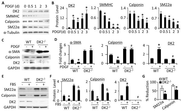
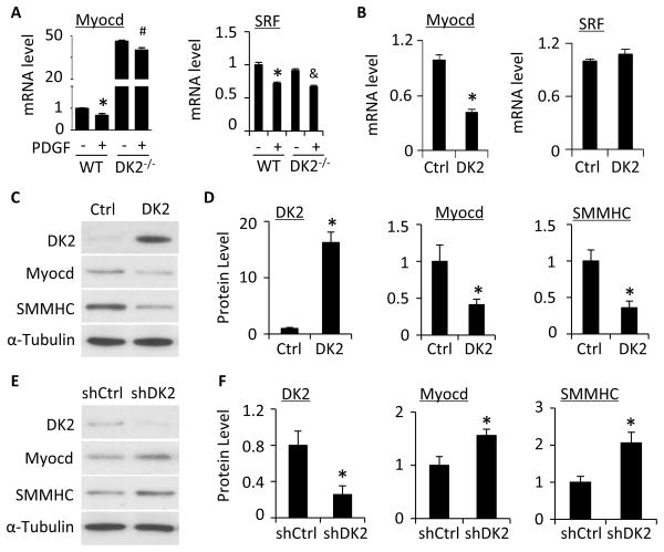
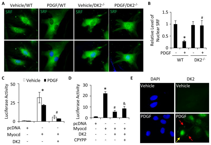
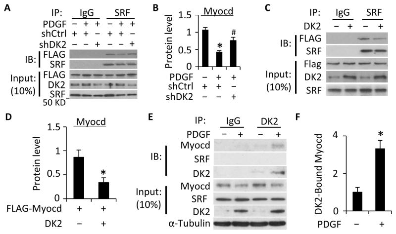
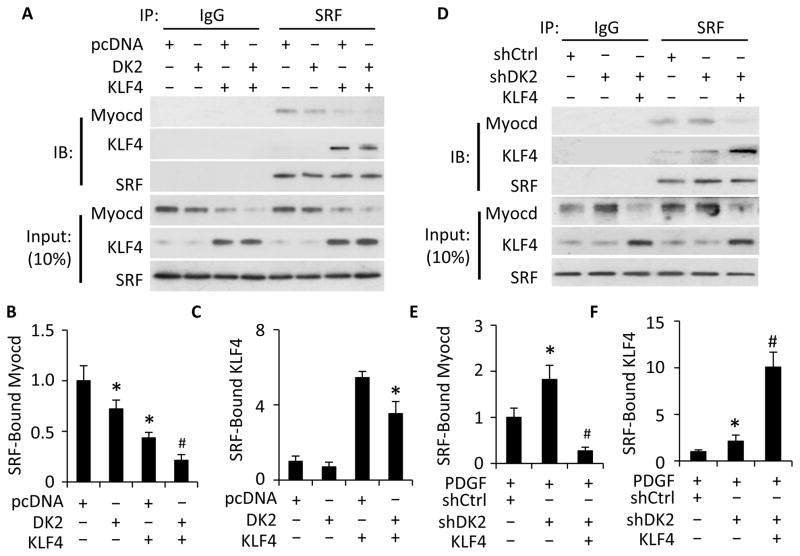
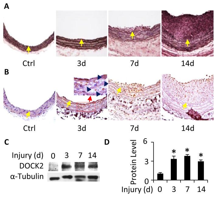
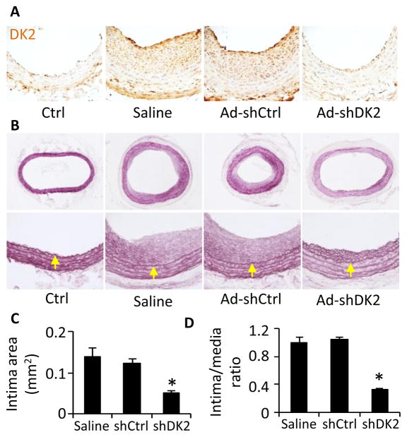
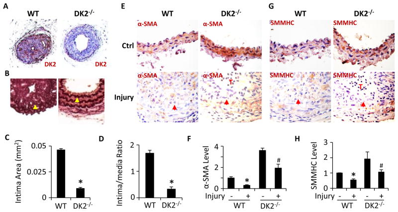
Methods and results: Platelet-derived growth factor-BB induced DOCK2 expression while modulating SMC phenotype. DOCK2 deficiency diminishes platelet-derived growth factor-BB or serum-induced downregulation of SMC markers. Conversely, DOCK2 overexpression inhibits SMC marker expression in primary cultured SMC. Mechanistically, DOCK2 inhibits myocardin expression, blocks serum response factor nuclear location, attenuates myocardin binding to serum response factor, and thus attenuates myocardin-induced smooth muscle marker promoter activity. Moreover, DOCK2 and Kruppel-like factor 4 cooperatively inhibit myocardin-serum response factor interaction. In a rat carotid artery balloon-injury model, DOCK2 is induced in media layer SMC initially and neointima SMC subsequently after vascular injury. Knockdown of DOCK2 dramatically inhibits the neointima formation by 60%. Most importantly, knockout of DOCK2 in mice markedly blocks ligation-induced intimal hyperplasia while restoring SMC contractile protein expression.

Conclusions: Our studies identified DOCK2 as a novel regulator for SMC phenotypic modulation and vascular lesion formation after vascular injury. Therefore, targeting DOCK2 may be a potential therapeutic strategy for the prevention of vascular remodeling in proliferative vascular diseases.

Keywords: cell proliferation; dedicator of cytokinesis 2; vascular remodeling.


Figures

Figure 1
Figure 1. DOCK2 mediated SMC phenotypic modulation
A, Time-dependent induction of DOCK2 (DK2) and SMC markers by PDGF-BB (10 ng/ml). B, Quantitative analysis of protein expression shown in A by normalizing to α-Tubulin. *, P<0.05 vs vehicle-treated group (0 h). n=3. C, Knockout of DOCK2 blocked PDGF-BB-induced downregulation of SMC marker proteins. SMCs isolated from wild type (WT) and DOCK2 knockout (DK2−/−) mice were treated with vehicle (−) or PDGF-BB (+) for 36 h. DK2, α-SMA, and calponin protein expression was detected by western blot. D, Protein levels shown in C were quantified by normalizing to GAPDH. Fold changes are shown. *, P<0.05 vs vehicle-treated WT group (−); #, P>0.05 vs vehicle-treated DK2−/− group, n=3. E, Serum (FBS)-induced SMC phenotypic modulation in WT and DK2−/− SMCs. Cells were treated with vehicle (−) or 10% FBS (+) for 48 h. SM22α, calponin, and DK2 protein expression was detected by western blot. F, Protein levels shown in E were quantified by normalizing to GAPDH. *, P<0.05 vs vehicle-treated WT group (−); &, P<0.05 vs vehicle-treated DK2−/− group; #, P>0.05 vs vehicle-treated DK2−/− group. n=3. G, DOCK2−/− attenuated FBS-induced SMC phenotypic modulation. The percentage downregulation of SMC markers by FBS was calculated by the following formula: (untreated value − FBS-treated)/untreated value)×100. *, P<0.05 vs WT group for each corresponding marker, n=3.
Figure 2
Figure 2. DOCK2 modulated SMC phenotype through downregulating myocardin
A, Knockout of DOCK2 (DK2−/−) increased myocardin (Myocd) but not SRF mRNA expression. SMCs isolated from WT or DK2−/− mice were treated with vehicle (−) or PDGF-BB (+) for 24 h. Myocd and SRF mRNA expression was detected by qPCR. *, P<0.05 vs vehicle-treated (−) WT group, #, P>0.05 vs vehicle-treated DK2−/− group. &, P<0.05 vs vehicle-treated DK2−/− group. n=3. B, Ectopic expression of DK2 inhibited Myocd but not SRF mRNA expression. Myocd and SRF mRNA expression was detected by qPCR. *, P<0.05 compared with control plasmid group (Ctrl), n=3. C, Ectopic expression of DK2 blocked Myocd and SMC marker protein expression. The protein expression of DK2, Myocardin, and SMMHC was detected by western blot. D, Quantification of protein levels shown in C by normalized to α-Tubulin. *, P<0.05 compared with Ctrl group, n=3. E, Knockdown of DK2 by shRNA enhanced Myocd and SMMHC protein expression in SMC. Primary cultured SMCs were transfected with control (shCtrl) or DOCK2 shRNA (shDK2) followed by PDGF-BB-treatment. The protein expression of DK2, Myocd, and SMMHC was detected by western blot. F, Quantification of protein levels shown in E by normalized to α-Tubulin. *, P<0.05 compared with shCtrl group, n=3.
Figure 3
Figure 3. DOCK2 blocked SRF nuclear translocation and myocardin-mediated SMC marker gene transcription
A, DOCK2 deficiency (DK2−/−) restored PDGF-BB-blocked SRF nuclear location. WT and DK2−/− SMCs were treated with vehicle or PDGF-BB for 24 h. SRF cellular location was detected by immunostaining. DAPI stained the nuclei. B, Quantification of nuclear SRF level shown in A by normalizing to the signal intensity in vehicle-treated WT SMCs (set as 1). *, P<0.05 vs vehicle-treated WT SMCs (−); #, P>0.05 vs vehicle-treated DK2−/− SMCs (−). n=3. C, DK2 blocked Myocd-activated α-SMA promoter activity. pcDNA, Myocd, or DK2 plasmid were co-transfected with α-SMA promoter as indicated into SMCs followed by vehicle or PDGF-BB treatment for 24 h. Luciferase assay was performed. *, P<0.05 vs pcDNA-transfected groups; #, P<0.05 vs Myocd alone-transfected groups. n=3. D, DK2 blocked α-SMA promoter activity independent of Rac. pcDNA, Myocd, or DK2 plasmid were co-transfected with α-SMA promoter into SMCs followed by vehicle (−) or CPYPP (25 μM) treatment as indicated for 24 h. Luciferase assay was performed. *, P<0.05 vs pcDNA-transfected group; #, P<0.05 vs Myocd alone-transfected group. &, P>0.05 vs Myocd/DK2 co-transfected group (n=3). E, DOCK2 nuclear location. DOCK2 cellular location was detected by immunostaining. DK2 was located on both cytoplasm membrane (red arrows) and nuclei (yellow arrow) of SMCs upon PDGF-BB treatment in addition to the cytoplasm.
Figure 4
Figure 4. DOCK2 inhibited Myocd-SRF interaction
A, Knockdown of DOCK2 (DK2) restored PDGF-BB-blocked Myocardin-SRF interaction. SMCs were transduced with scramble (shCtrl) or DK2 shRNA (shDK2) and transfected with Flag-tagged Myocd cDNA as indicated. Cells were then treated with PDGF-BB. Cell lysates were immunoprecipitated (IP) with normal IgG or SRF antibody followed by immunobloting (IB) with Flag or SRF antibody. B, Quantification of SRF-bound Myocd by normalizing to the input Myocd level in each treatment and set the vehicle-treated group as 1. *, P<0.05 vs the vehicle treated group (−); #, P<0.05 vs PDGF-BB and shCtrl-treated groups (n=3). C. DK2 overexpression suppressed Myocardin-SRF interaction. Control (−) or DK2 expression plasmids were co-transfected with Flag-tagged Myocd into SMCs followed by Co-IP with IgG or SRF antibody and IB with Flag antibody. D, SRF-bound Myocd was quantified similarly as in B. *, P<0.05 vs control plasmid-transfected group (−), n=3. E, Co-IP with endogenous proteins indicated that Myocd physically interacted with DK2. Primary cultured SMCs were treated with vehicle (−) or PDGF-BB (+) for 24 h. Cell lysates were Co-IP with normal IgG or DK2 antibody, and blotted with Myocd or SRF antibody as indicated. Myocd-DK2 interaction was enhanced by PDGF-BB. F, Quantification of DK2-bound Myocd shown in E. *, P<0.05 vs vehicle-treated group, n=3.
Figure 5
Figure 5. DOCK2 and KLF4 cooperatively inhibited Myocd-SRF binding
A, Co-expression of DOCK2 (DK2) and KLF4 completely blocked Myocd-SRF binding. SMCs were co-transfected with pcDNA, DK2, and/or KLF4 expression plasmids as indicated followed by serum starvation for 24 h. Cell lysates were immunoprecipitated (IP) with IgG and SRF antibody. The immunoprecipitates were blotted (IB) with Myocd, KLF4 and SRF antibody as indicated. B, Quantification of SRF-bound Myocd in A. *, P<0.05 vs pcDNA-transfected group (set as 1); #, P<0.05 vs all other groups (n=3). C, Quantification of SRF-bound KLF4 in A. *, P<0.05 vs pcDNA/KLF4 plasmid-transfected group, n=3. D, KLF4 blocked DOCK2 knockdown-enhanced Myocd-SRF binding. SMCs were transfected with control (shCtrl) or DK2 shRNA (shDK2) for 48 h followed by transfection of pcDNA (−) or KLF4 expression plasmid (+) as indicated and PDGF-BB treatment for 24 h. Cell lysates were Co-IP with IgG and SRF antibody. The precipitated proteins were blotted (IB) with Myocd, KLF4 and SRF antibody as indicated. E, Quantification of SRF-bound Myocd in D. *, P< 0.05 compared with shCtrl group (set as 1); #, P<0.05 vs shDK2-treated cells without KLF4 plasmid (n=3). F. Quantification of SRF-bound KLF4 in D. *, P< 0.05 compared with shCtrl group (set as 1); #, P<0.05 compared other two groups (n=3).
Figure 6
Figure 6. Balloon injury induced DOCK2 expression in tunica media and neointima SMCs
A, Balloon injury induced progressive neointima formation. Rat left carotid arteries were injured for 3, 7, and 14 days as indicated. The right non-injured carotid arteries were used as controls (Ctrl). Artery sections were stained with Elastica van Gieson solution. Yellow arrows indicate the internal elastin lamina. B, DOCK2 was induced in tunica media initially and neointima VSMCs subsequently following injury. Artery sections were incubated with DOCK2 antibody followed by horseradish peroxidase-conjugated secondary antibody and DAB staining. Dark blue arrows in the enlarged image of the red box in the 3d sections show representative SMCs expressing DOCK2. Yellow arrows indicate the internal elastin lamina. C, DOCK2 expression in injured arteries was detected by western blot. Data shown are a representative result of 3 independent experiments. D, Quantification of DOCK2 expression by normalizing to α-Tubulin. *, P<0.05 compared to uninjured arteries (0 d).
Figure 7
Figure 7. Knockdown of DOCK2 blocked neointima formation
A, DOCK2 expression was efficiently blocked by adenovirus delivery of DOCK2 shRNA. Immediately after balloon injury, the injured rat carotid arteries were incubated with sterile saline solution, adenovirus expressing scramble (Ad-shCtrl), or DOCK2 shRNA (Ad-shDK2) as indicated. 14 days later, artery sections were stained with DOCK2 antibody. DOCK2 (DK2) expression was visualized by DAB staining. B, DOCK2 knockdown blocked injury-induced neointima formation. Artery sections were stained with Elastica van Gieson solution. Yellow arrows indicate internal elastic lamina. C and D, Quantification of intima area and intima/media ratio. *, P<0.05 compared with saline- or Ad-shCtrl-treated arteries (n=6).
Figure 8
Figure 8. DOCK2 deficiency attenuated injury-induced neointima formation and SMC phenotypic modulation in mouse arteries
A, Ligation injury induced DOCK2 expression in wild type (WT), but not DOCK2 knockout (DK2−/−) mouse carotid arteries. Mouse left carotid arteries were injured by ligation for 28 days. DOCK2 (DK2) expression was detected by immunohistochemistry (IHC) staining using DK2 antibody and DAB visulization. B, DK2 knockout blocked ligation-induced neointima formation. Artery sections were stained with Elastica van Gieson solution. Arrows indicate internal elastic lamina. C-D, Quantification of intima area (C) and intima/media ratio (D) of the injured arteries. *, P<0.05 vs WT mouse carotid arteries with ligation (n=6). E-H, DK2−/− increased α-SMA (E) and SMMHC (G) expression in both control (Ctrl) and ligation-injured arteries. α-SMA and SMMHC was detected by IHC staining using α-SMA (E) and SMMHC (G) antibodies followed by DAB visualization. Arrows indicate internal elastic lamina. T indicates the thrombus in injured DK2−/− arteries. α-SMA (F) and SMMHC (H) expression was quantified by normalizing to the DAB intensity in the WT Ctrl arteries (set as 1). *, P<0.05 vs WT arteries without injury (−); #, P<0.05 vs WT arteries with injury for each individual proteins (n=6).

References

    1. Schwartz SM, deBlois D, O’Brien ER. The intima. Soil for atherosclerosis and restenosis. Circulation research. 1995;77:445–465. - PubMed
    1. Neitzel GF, Barboriak JJ, Pintar K, Qureshi I. Atherosclerosis in aortocoronary bypass grafts. Morphologic study and risk factor analysis 6 to 12 years after surgery. Arteriosclerosis. 1986;6:594–600. - PubMed
    1. Schwartz SM. Smooth muscle migration in atherosclerosis and restenosis. The Journal of clinical investigation. 1997;100:S87–89. - PubMed
    1. Dzau VJ, Braun-Dullaeus RC, Sedding DG. Vascular proliferation and atherosclerosis: New perspectives and therapeutic strategies. Nature medicine. 2002;8:1249–1256. - PubMed
    1. Owens GK, Kumar MS, Wamhoff BR. Molecular regulation of vascular smooth muscle cell differentiation in development and disease. Physiological reviews. 2004;84:767–801. - PubMed

Publication types

MeSH terms