. 2015 Oct;103(10):3212-25.
doi: 10.1002/jbm.a.35449. Epub 2015 Mar 31.

Porous three-dimensional carbon nanotube scaffolds for tissue engineering

Affiliations

Porous three-dimensional carbon nanotube scaffolds for tissue engineering

Gaurav Lalwani et al. J Biomed Mater Res A. 2015 Oct.

Abstract

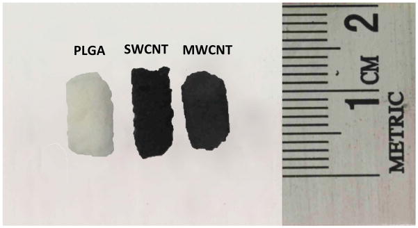
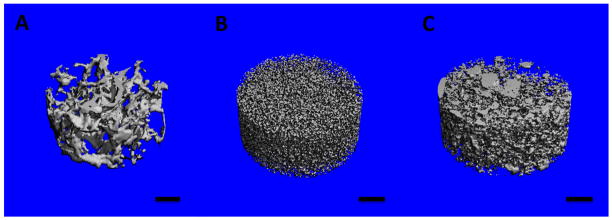
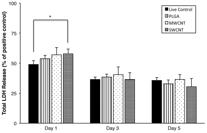
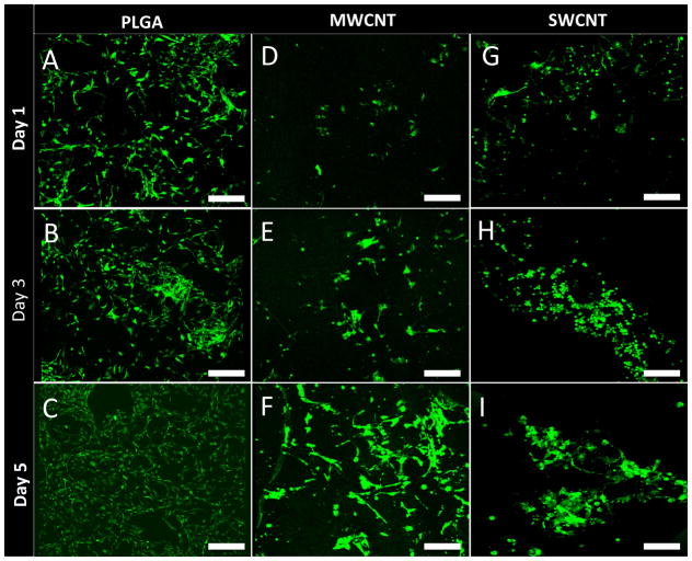
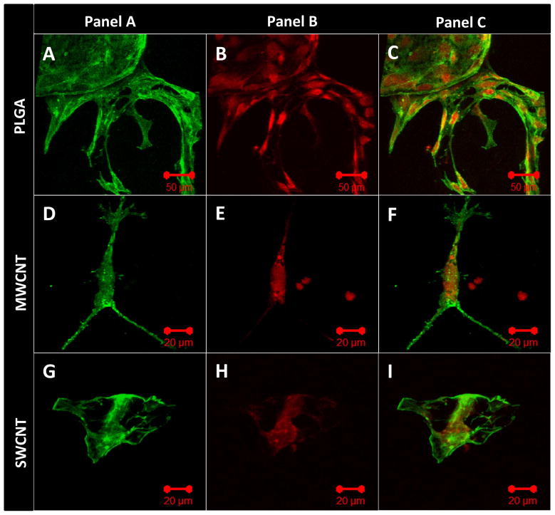
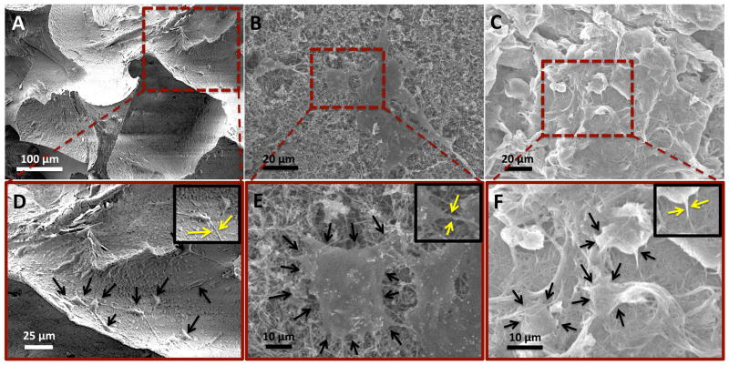
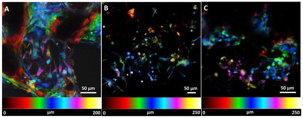
Assembly of carbon nanomaterials into three-dimensional (3D) architectures is necessary to harness their unique physiochemical properties for tissue engineering and regenerative medicine applications. Herein, we report the fabrication and comprehensive cytocompatibility assessment of 3D chemically crosslinked macrosized (5-8 mm height and 4-6 mm diameter) porous carbon nanotube (CNT) scaffolds. Scaffolds prepared via radical initiated thermal crosslinking of single- or multiwalled CNTs (SWCNTs and MWCNTs) possess high porosity (>80%), and nano-, micro-, and macroscale interconnected pores. MC3T3 preosteoblast cells on MWCNT and SWCNT scaffolds showed good cell viability comparable to poly(lactic-co-glycolic) acid (PLGA) scaffolds after 5 days. Confocal live cell and immunofluorescence imaging showed that MC3T3 cells were metabolically active and could attach, proliferate, and infiltrate MWCNT and SWCNT scaffolds. SEM imaging corroborated cell attachment and spreading and suggested that cell morphology is governed by scaffold surface roughness. MC3T3 cells were elongated on scaffolds with high surface roughness (MWCNTs) and rounded on scaffolds with low surface roughness (SWCNTs). The surface roughness of scaffolds may be exploited to control cellular morphology and, in turn, govern cell fate. These results indicate that crosslinked MWCNTs and SWCNTs scaffolds are cytocompatible, and open avenues toward development of multifunctional all-carbon scaffolds for tissue engineering applications.

Keywords: carbon nanotubes; cytotoxicity; scaffolds; three dimensional; tissue engineering.


Figures

Figure 1
Figure 1
Optical images of representative three-dimensional porous poly(lactic-co-glycolic) acid, single walled carbon nanotube and multi walled carbon nanotube scaffolds prepared as cylinders (5 mm diameter, ~8–10 mm length).
Figure 2
Figure 2
Representative scanning electron microscopy images of (A) poly(lactic-co-glycolic) acid, (B) multi walled carbon nanotube and (C) single walled carbon nanotube scaffolds. Yellow arrows images (B) and (C) correspond to the formation of nanoscale junctions (crosslinks) between CNTs.
Figure 3
Figure 3
Representative three-dimensional microcomputed tomography reconstructions of subsections of (A) poly(lactic-co-glycolic) acid, (B) multi walled carbon nanotube and (C) single walled carbon nanotube scaffolds. The blue color represents void spaces. Scale bars are 200 μm.
Figure 4
Figure 4
Cytotoxicity evaluation using LDH assay after 1, 3, and 5 days of MC3T3 cell culture on poly(lactic-co-glycolic) acid, multi walled carbon nanotube, and single walled carbon nanotube scaffolds. Chart represents total LDH release (%) normalized to positive controls (100% dead cells). Data is represented as means ± standard deviation. Groups with a significant difference (p < 0.05) are marked with “*”.
Figure 5
Figure 5
Representative calcein-AM stained green fluorescence images of MC3T3 cells on (A–C) poly(lactic-co-glycolic) acid, (D–F) multi walled carbon nanotube and (G–I) single walled carbon nanotube scaffolds after 1, 3, and 5 days of culture. Presence of live cells (green fluorescence) on all scaffold groups can be observed. Scale bars are 200 μm.
Figure 6
Figure 6
Representative immunofluorescence images of MC3T3 cells cultured on (A–C) poly(lactic-co-glycolic) acid, (D–F) multi walled carbon nanotube and (G–I) single walled carbon nanotube scaffolds after 1 day stained green (calceinAM) for actin cytoskeleton (Panel A) and red (rhodamine conjugated mAb) for focal adhesions, i.e. vinculin protein (Panel B). Panel C shows superimposed images of panels A and B showing the co-localization of actin filaments and vinculin protein.
Figure 7
Figure 7
Representative immunofluorescence images of MC3T3 cells cultured on (A–C) poly(lactic-co-glycolic) acid, (D–F) multi walled carbon nanotube and (G–I) single walled carbon nanotube scaffolds after 5 days stained green (calceinAM) for actin cytoskeleton (Panel A) and red (rhodamine conjugated mAb) for cell proliferation marker, i.e. Ki-67 protein (Panel B). Panel C shows superimposed images of panels A and B showing the co-localization of actin filaments and Ki-67 protein.
Figure 8
Figure 8
Representative SEM images showing adhesion of MC3T3 cells on (A and D) poly(lactic-co-glycolic) acid, (B and E) multi walled carbon nanotube, and (C and F) single walled carbon nanotube scaffolds. Formation of cytoplasmic extensions (filopodia and pseudopodia) can be observed for each scaffold group (inset in images D, E and F).
Figure 9
Figure 9
Representative spectrally color coded images of calcein-AM stained MC3T3 cells a function of confocal Z-depth (i.e. cellular infiltration) after 5 days of culture on (A) poly(lactic-co-glycolic) acid, (B) multi walled carbon nanotube and (C) single walled carbon nanotube scaffolds. Presence of cells can be detected upto a depth of ~200–300 μm for each scaffold group.

References

    1. Discher DE, Mooney DJ, Zandstra PW. Growth factors, matrices, and forces combine and control stem cells. Science. 2009;324:1673–7. - PMC - PubMed
    1. Engler AJ, Sen S, Sweeney HL, Discher DE. Matrix elasticity directs stem cell lineage specification. Cell. 2006;126:677–89. - PubMed
    1. Dalby MJ, Gadegaard N, Oreffo RO. Harnessing nanotopography and integrin-matrix interactions to influence stem cell fate. Nature materials. 2014;13:558–69. - PubMed
    1. Hui EE, Bhatia SN. Micromechanical control of cell-cell interactions. Proceedings of the National Academy of Sciences of the United States of America. 2007;104:5722–6. - PMC - PubMed
    1. Peltola SM, Melchels FP, Grijpma DW, Kellomaki M. A review of rapid prototyping techniques for tissue engineering purposes. Annals of medicine. 2008;40:268–80. - PubMed

Publication types

Substances

LinkOut - more resources