

Characterization of PD-L1 Expression and Associated T-cell Infiltrates in Metastatic Melanoma Samples from Variable Anatomic Sites
- PMID: 25788491
- PMCID: PMC4490112
- DOI: 10.1158/1078-0432.CCR-14-3073
Characterization of PD-L1 Expression and Associated T-cell Infiltrates in Metastatic Melanoma Samples from Variable Anatomic Sites
Abstract
Purpose: Programmed death ligand-1 (PD-L1) tumor expression represents a mechanism of immune escape for melanoma cells. Drugs blocking PD-L1 or its receptor have shown unprecedented activity in melanoma, and our purpose was to characterize tumor PD-L1 expression and associated T-cell infiltration in metastatic melanomas.
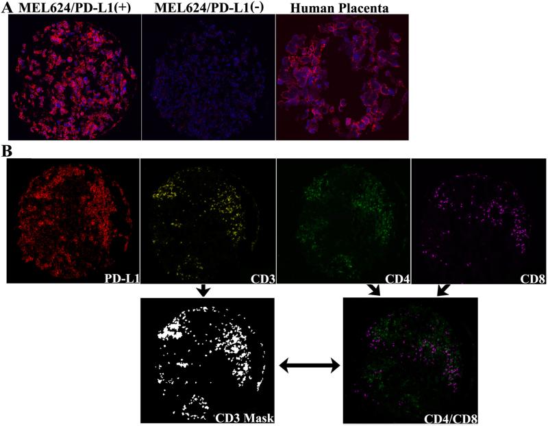
Experimental design: We used a tissue microarray (TMA) consisting of two cores from 95 metastatic melanomas characterized for clinical stage, outcome, and anatomic site of disease. We assessed PD-L1 expression and tumor-infiltrating lymphocyte (TIL) content (total T cells and CD4/CD8 subsets) by quantitative immunofluorescence.
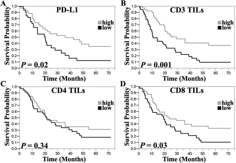
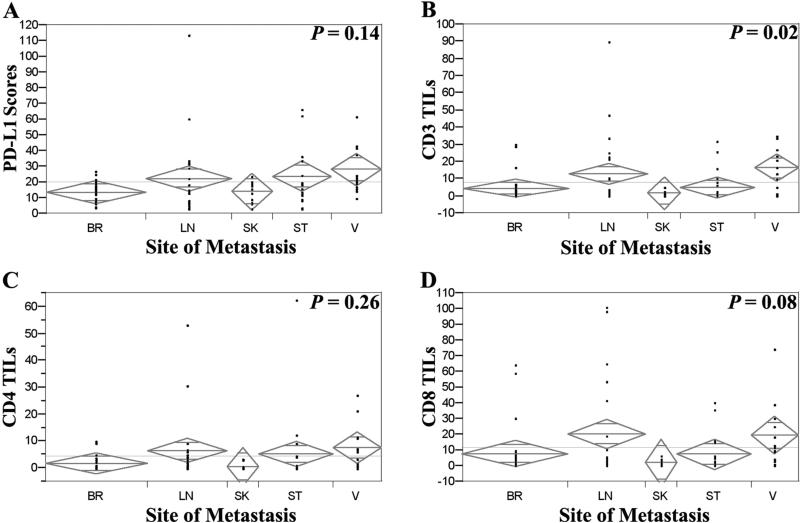
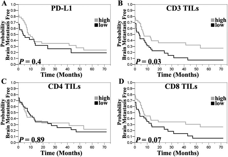
Results: High PD-L1 expression was associated with improved survival (P = 0.02) and higher T-cell content (P = 0.0005). Higher T-cell content (total and CD8 cells) was independently associated with improved overall survival; PD-L1 expression was not independently prognostic. High TIL content in extracerebral metastases was associated with increased time to developing brain metastases (P = 0.03). Cerebral and dermal metastases had slightly lower PD-L1 expression than other sites, not statistically significant. Cerebral metastases had less T cells (P = 0.01).
Conclusions: T-cell-infiltrated melanomas, particularly those with high CD8 T-cell content, are more likely to be associated with PD-L1 expression in tumor cells, an improved prognosis, and increased time to development of brain metastases. Studies of T-cell content and subsets should be incorporated into trials of PD-1/PD-L1 inhibitors to determine their predictive value. Furthermore, additional studies of anatomic sites with less PD-L1 expression and T-cell infiltrate are needed to determine if discordant responses to PD-1/PD-L1 inhibitors are seen at those sites.
©2015 American Association for Cancer Research.
Figures
References
-
- Acquavella N, Kluger H, Rhee J, Farber L, Tara H, Ariyan S, et al. Toxicity and activity of a twice daily high-dose bolus interleukin 2 regimen in patients with metastatic melanoma and metastatic renal cell cancer. J Immunother. 2008;31:569–76. - PubMed
-
- Miller RL, Steis RG, Clark JW, Smith JW, 2nd, Crum E, McKnight JE, et al. Randomized trial of recombinant alpha 2b-interferon with or without indomethacin in patients with metastatic malignant melanoma. Cancer Res. 1989;49:1871–6. - PubMed
-
- Sznol M. Blockade of the B7-H1/PD-1 pathway as a basis for combination anticancer therapy. Cancer J. 2014;20:290–5. - PubMed
Publication types
MeSH terms
Substances
Grants and funding
LinkOut - more resources
Full Text Sources
Other Literature Sources
Medical
Research Materials
