Review
. 2015 Apr;28(2):337-418.
doi: 10.1128/CMR.00117-14.

The challenge of efflux-mediated antibiotic resistance in Gram-negative bacteria

Affiliations
Review

The challenge of efflux-mediated antibiotic resistance in Gram-negative bacteria

Xian-Zhi Li et al. Clin Microbiol Rev. 2015 Apr.

Abstract

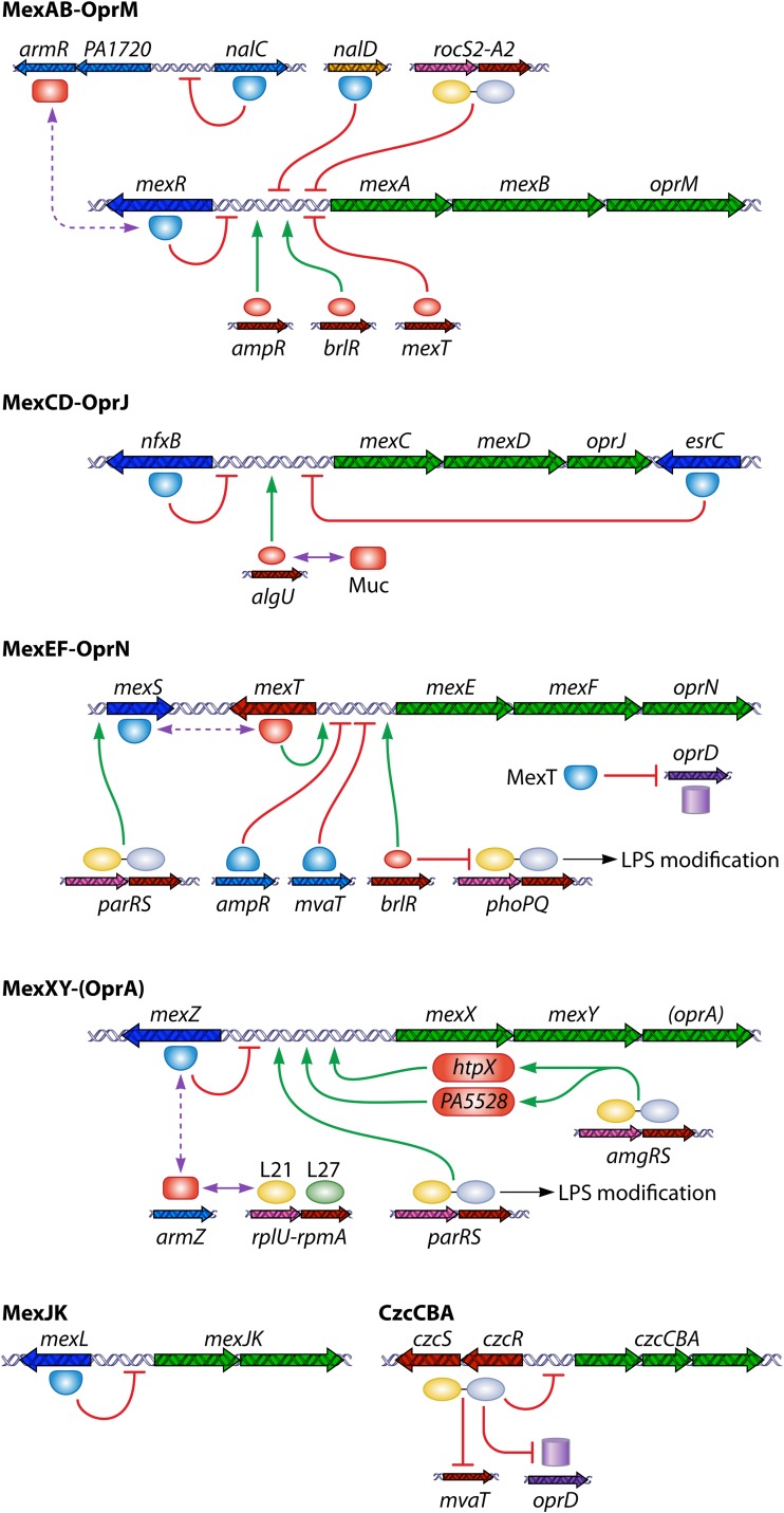
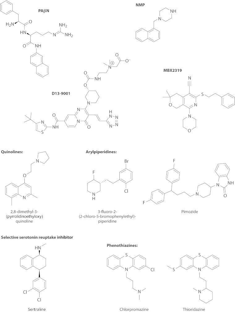
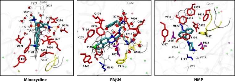
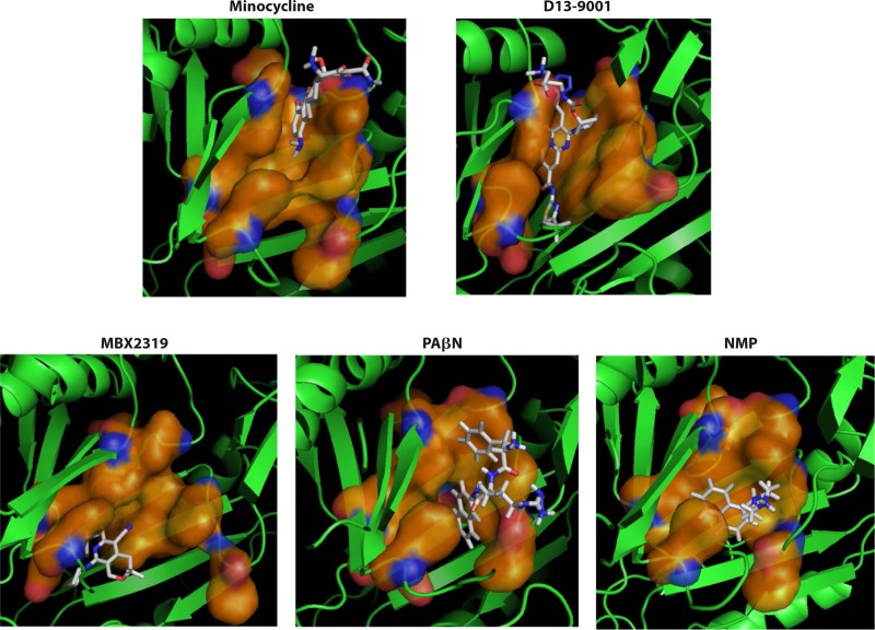
The global emergence of multidrug-resistant Gram-negative bacteria is a growing threat to antibiotic therapy. The chromosomally encoded drug efflux mechanisms that are ubiquitous in these bacteria greatly contribute to antibiotic resistance and present a major challenge for antibiotic development. Multidrug pumps, particularly those represented by the clinically relevant AcrAB-TolC and Mex pumps of the resistance-nodulation-division (RND) superfamily, not only mediate intrinsic and acquired multidrug resistance (MDR) but also are involved in other functions, including the bacterial stress response and pathogenicity. Additionally, efflux pumps interact synergistically with other resistance mechanisms (e.g., with the outer membrane permeability barrier) to increase resistance levels. Since the discovery of RND pumps in the early 1990s, remarkable scientific and technological advances have allowed for an in-depth understanding of the structural and biochemical basis, substrate profiles, molecular regulation, and inhibition of MDR pumps. However, the development of clinically useful efflux pump inhibitors and/or new antibiotics that can bypass pump effects continues to be a challenge. Plasmid-borne efflux pump genes (including those for RND pumps) have increasingly been identified. This article highlights the recent progress obtained for organisms of clinical significance, together with methodological considerations for the characterization of MDR pumps.


Figures

FIG 1
FIG 1
Location of drug efflux pumps and pathways of drug influx and efflux across the OM and IM in Gram-negative bacteria. The influx of drugs (shown as pills) through the OM occurs in one or more of the following three pathways: porin channels (e.g., OmpF of E. coli and OprF of P. aeruginosa), specific protein channels (e.g., CarO of A. baumannii and OprD of P. aeruginosa for carbapenems), and the LPS-containing asymmetric lipid bilayer region. After their entry into the periplasmic space, the drug molecules can further penetrate the IM via diffusion. However, these drugs can be extruded out of the cell by efflux transporters, which exist as either single-component pumps (“singlet”; e.g., Tet pumps) or multicomponent pumps (e.g., AcrAB-TolC and MexAB-OprM tripartite efflux systems that each typically contain a pump, an OM channel protein [OMP], and an accessory membrane fusion protein [MFP]). While the singlet pumps may take up the drug from the cytosol and the periplasm and function with porins or other types of protein channels to make the efflux process effective, the multicomponent exporters capture their substrates from the periplasm and the IM and directly pump them into the medium. The competition between the influx and efflux processes ultimately determines the steady state of drug molecules in bacterial cells. With the lipophilic drug molecules that cross the OM slowly or the hydrophilic drugs that penetrate the A. baumannii/P. aeruginosa low-permeability porins (i.e., “slow porins”), the efflux mechanism become very effective, thus being able to yield MDR. In contrast, with the less hydrophobic and smaller drug molecules that can rapidly penetrate, for example, E. coli porins, efflux is not effective to counteract drug influx, thus hardly decreasing the concentrations of the drug in the cell.
FIG 2
FIG 2
Drug transport mechanism of AcrB. Shown is the asymmetric crystal structure of AcrB (Protein Data Bank accession number 2DRD), viewed from outside the cell, with the top portion cut off for clarity. Conformational cycling of 3 AcrB protomers, in access (blue), binding (red), and extrusion (green), is seen by cocrystallization of AcrB with its substrate minocycline, shown in a yellow stick model.
FIG 3
FIG 3
Interaction of drug substrates and the AcrB-binding protomer analyzed with Autodock Vina docking software. Substrates are shown to bind to either the upper part (groove binder) (doxorubicin [A] and tetracycline [B]) or the lower part (cave binder) (chloramphenicol [C] and cyclohexane [D]) of the distal binding site. (Modified from reference .)
FIG 4
FIG 4
Regulation of expression of the AcrAB-TolC efflux system of E. coli. Transcription of the acrAB and tolC genes is not genetically clustered but is often regulated by common regulators at multiple levels. The local repressor AcrR represses acrAB expression directly. Other regulators include the AcrS repressor of the AcrEF system, histone-like nucleoid structuring protein (H-NS), and the SdiA global regulator. Three global regulators, MarA, SoxS, and Rob, positively control the expression of acrAB, tolC, and micF. The micF transcript inhibits the translation of OmpF porin mRNA. The two-component regulatory systems EvgAS and/or PhoQP can enhance acrAB and tolC expression. The red lines show the repression of the transcription of the relevant gene by the repressors AcrR, AcrS, H-NS, MarR, and SoxR. The green arrowed lines reveal the activation of relevant gene expression by the activators MarA, SoxS, SdiA, Rob, EvgS, and PhoP (SoxS and Rob can also stimulate MarA expression). MarB modulates MarA expression. Several regulators can bind with certain ligands (such as antimicrobial agents and metabolites) or be induced by oxidative stress and thus become inactivated (in the case of AcrR, MarR, and SoxR) or activated (in the case of Rob when binding with bile salts or fatty acids). Mutational changes can lead to the inactivation of AcrR, AcrS, MarR, and SoxR. The crystal structures of AcrR, MarA, MarR, SoxR, and Rob are available with identified ligand-binding domains and conformational changes for regulation. Regulation of AcrAB by noncoding RNAs has also been identified (see the text). Overall, under various conditions, these multiple regulation mechanisms can together produce MDR by allowing simultaneously decreased influx (via OmpF porin) and increased efflux (via AcrAB-TolC) of antimicrobial agents, which can be captured by the pump complex from the outer and/or inner leaflets of the IM and the periplasm (but not directly from the cytosol). Expression of acrZ (whose product can be copurified with AcrB) is coregulated with that of acrAB via MarA, SoxS, and Rob.
FIG 5
FIG 5
Regulation of expression of RND multidrug efflux systems of P. aeruginosa. Of 12 RND pump operons identified in this organism, half of them (presented in green, with the arrows showing their transcriptional directions) are regulated under a local regulator (mostly by a repressor [MexR, NfxB, EsrC, MexZ, or MexL] or by an activator [MexT] encoded by a gene adjacent to the efflux operons) or a two-component system of CzcRS for the czcCBA operon. (RND pump operons with no identified local regulatory genes are not included.) The red lines show the repression of the transcription of the relevant gene by repressors, while the green arrows reveal positive regulation by the regulators. Local repressors are controlled by antirepressor proteins (ArmR and ArmZ) and can also bind to ligands (e.g., antimicrobial agents or metabolites, including quorum-sensing molecules) or be induced under various conditions (oxidative or nitrosative stress or the presence of different agents). The ribosomal proteins L21 and L27, encoded by rplU-rpmA, indirectly upregulate ArmZ and negatively control mexXY expression. Activation of mexXY expression also occurs through positive control by the AmgRS two-component system via the HtpX and PA5528 proteins. Additional regulators encoded by the genes not genetically clustered with the efflux operons also participate in regulation. Mutational changes can also lead to an inactivation of regulators (ArmR, ArmZ, MexR, NalC, NalD, MexS, and MexZ). The crystal structures of MexR and MexZ are available. AlgU is a sigma factor required for the oxidative stress response, and its activity is controlled by Muc, the inner membrane-associated proteins involved in the production of alginate exopolysaccharide and with their encoding genes clustered with algU. Some regulators, including AmpR, BrlR, MvaT, ParRS, and RocS2-RocA2, are involved in the regulation of expression of other genes, e.g., downregulation of OprD by MexT and CzcR (in carbapenem resistance) or LPS modification by BrlR and ParR (affecting polymyxin susceptibility). Certain gene products (e.g., BrlR) are involved in gene regulation in biofilm cells. See the text for detail.
FIG 6
FIG 6
Chemical structures of RND pump inhibitors.
FIG 7
FIG 7
MD simulation-predicted binding of the inhibitors PAβN and NMP, with the substrate minocycline shown as a reference. The positions of ligands initially predicted by docking (Autodock Vina) are shown as thin gray sticks, and those in the final phase of MD simulation are shown as thick blue sticks. AcrB residues within 3.5 Å of the ligand are shown in stick models (red, green, or yellow, if they belong to the distal pocket, proximal pocket, or G-loop, respectively). For NMP, two somewhat different equilibrium positions were obtained, and only one is shown here. (Modified from reference .)
FIG 8
FIG 8
Binding of various inhibitors determined by MD simulation. Although the binding of PAβN and NMP was examined previously (93), the simulation process was extended to >300 ns. The orange surface shows the distal binding pocket (defined previously [91]), and the inhibitor molecules are shown in sticks with CPK colors. AcrB is shown in green cartoon models, and the part closer to the viewer was removed for clarity. This figure was drawn by using the program Pymol, on the basis of data reported previously (96).
None
None
None

References

    1. World Health Organization. 2014. Antimicrobial resistance: global report on surveillance. World Health Organization, Geneva, Switzerland.
    1. D'Costa VM, King CE, Kalan L, Morar M, Sung WW, Schwarz C, Froese D, Zazula G, Calmels F, Debruyne R, Golding GB, Poinar HN, Wright GD. 2011. Antibiotic resistance is ancient. Nature 477:457–461. doi:10.1038/nature10388. - DOI - PubMed
    1. Forsberg KJ, Patel S, Gibson MK, Lauber CL, Knight R, Fierer N, Dantas G. 2014. Bacterial phylogeny structures soil resistomes across habitats. Nature 509:612–616. doi:10.1038/nature13377. - DOI - PMC - PubMed
    1. Davies J, Davies D. 2010. Origins and evolution of antibiotic resistance. Microbiol Mol Biol Rev 74:417–433. doi:10.1128/MMBR.00016-10. - DOI - PMC - PubMed
    1. Finley RL, Collignon P, Larsson DG, McEwen SA, Li X-Z, Gaze WH, Reid-Smith R, Timinouni M, Graham DW, Topp E. 2013. The scourge of antibiotic resistance: the important role of the environment. Clin Infect Dis 57:704–710. doi:10.1093/cid/cit355. - DOI - PubMed

Publication types

MeSH terms

Substances