. 2015 Nov;26(11):2765-76.
doi: 10.1681/ASN.2013101040. Epub 2015 Mar 18.

Akt Substrate of 160 kD Regulates Na+,K+-ATPase Trafficking in Response to Energy Depletion and Renal Ischemia

Affiliations

Akt Substrate of 160 kD Regulates Na+,K+-ATPase Trafficking in Response to Energy Depletion and Renal Ischemia

Daiane S Alves et al. J Am Soc Nephrol. 2015 Nov.

Abstract

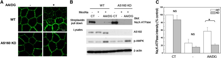
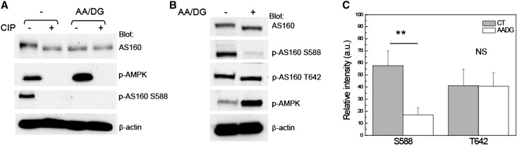
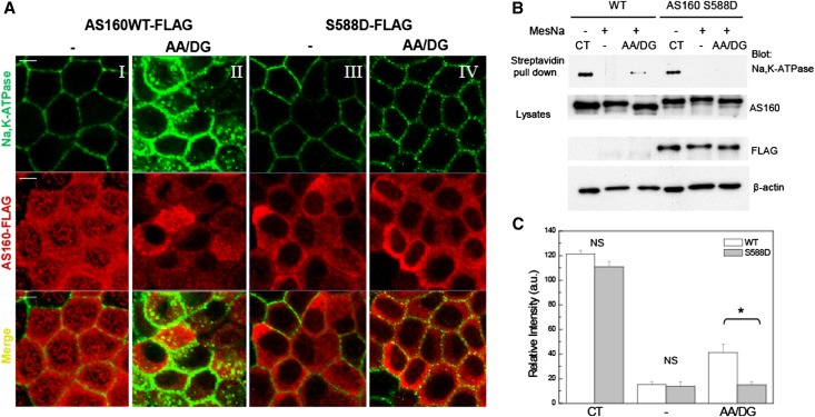
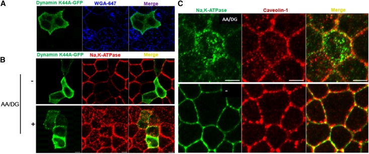
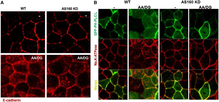
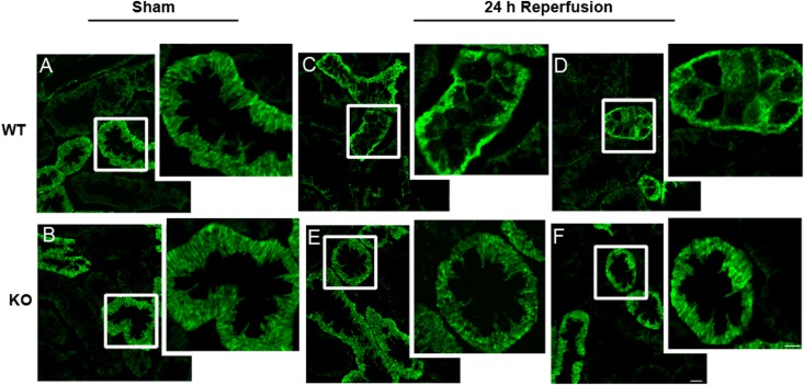
Renal ischemia and reperfusion injury causes loss of renal epithelial cell polarity and perturbations in tubular solute and fluid transport. Na(+),K(+)-ATPase, which is normally found at the basolateral plasma membrane of renal epithelial cells, is internalized and accumulates in intracellular compartments after renal ischemic injury. We previously reported that the subcellular distribution of Na(+),K(+)-ATPase is modulated by direct binding to Akt substrate of 160 kD (AS160), a Rab GTPase-activating protein that regulates the trafficking of glucose transporter 4 in response to insulin and muscle contraction. Here, we investigated the effect of AS160 on Na(+),K(+)-ATPase trafficking in response to energy depletion. We found that AS160 is required for the intracellular accumulation of Na(+),K(+)-ATPase that occurs in response to energy depletion in cultured epithelial cells. Energy depletion led to dephosphorylation of AS160 at S588, which was required for the energy depletion-induced accumulation of Na,K-ATPase in intracellular compartments. In AS160-knockout mice, the effects of renal ischemia on the distribution of Na(+),K(+)-ATPase were substantially reduced in the epithelial cells of distal segments of the renal tubules. These data demonstrate that AS160 has a direct role in linking the trafficking of Na(+),K(+)-ATPase to the energy state of renal epithelial cells.

Keywords: MDCK; cell biology; cell physiology; cell signaling; cell structure; epithelial; ischemia-reperfusion; transport physiology.


Figures

Figure 1.
Figure 1.
shRNA-mediated KD of AS160 inhibits the intracellular accumulation of Na,K-ATPase that is induced by energy depletion. (A) Immunofluorescence analysis is performed to detect the distribution of the endogenous Na,K-ATPase in MDCK renal epithelial cells in culture. MDCK cells are stained with an antibody directed against the sodium pump α-subunit (α5). WT MDCK cells and MDCK cells knocked down for AS160 expression (AS160 KD) are treated with (+) or without (−) AA/DG. Typical results from one of five experiments are shown. (B) Cell surface biotinylation. WT or AS160 KD cells are biotinylated at the basolateral surface, then subjected or not to energy depletion. The biotin that remained exposed at the cell surface after the treatments is stripped by treating the cells with MesNa. Biotinylated proteins are recovered through incubation with streptavidin agarose beads. The biotinylated Na,K-ATPase α-subunit is detected with the mAb α5. Lysates are blotted with anti-AS160, anti–phospho-AMPK antibody, and with anti–β-actin to assess total protein loading. The extent of energy depletion is assessed by probing for phospho-AMPK. (C) Quantification of the biotinylated Na,K-ATPase band intensity normalized to the β-actin levels. The results indicate that the fraction of the pool of sodium pump that was initially biotinylated at the cell surface that accumulated within the cell following energy depletion is lower in the AS160 KD cells compared with the WT line. *P<0.01 (n=4). CT, total biotinylated Na,K-ATPase in untreated cells; −, biotinylated Na,K-ATPase in untreated cells subjected to MesNa strip; AA/DG, biotinylated Na,K-ATPase in energy-depleted cells treated with MesNa; p-AMPK, phospho-AMPK. Bar, 5 μm.
Figure 2.
Figure 2.
The interaction between AS160 and Na,K-ATPase is not affected by energy depletion treatment. Endogenous Na,K-ATPase is immunoprecipitated using a mAb directed against the α-subunit (α5). Immunoprecipitates are probed with AS160 polyclonal antibody to detect endogenous AS160. MDCK cells are treated (+) or not (−) with energy depletion AA/DG, for 1 hour. (A) Immunoblot of the coimmunoprecipitation. Lysates are blotted with anti-AS160 to detect the endogenous levels of AS160 and with anti–β-actin as a control for the total protein loaded. (B) Coimmunoprecipitation quantification. The results indicate that the interaction between AS160 and sodium pump is not affected by energy depletion. n=3. IP, immunoprecipitation.
Figure 3.
Figure 3.
Energy depletion leads to AS160 dephosphorylation at S588. (A) The apparent molecular weight of AS160 decreases as a result of its dephosphorylation. MDCK cells are subjected (AA/DG) or not (−) to energy depletion for 1 hour. Total cell lysates are incubated in the presence (+) or absence (−) of 10 units of CIP for 30 minutes at 37°C. Lysates are blotted with anti-AS160 antibody to visualize the shift of the apparent molecular weight of AS160. Blots are also probed with anti–phospho-AMPK and p-AS160 S588 antibodies to document the efficacy of both the ATP depletion and the CIP treatments. Anti–β-actin is also used to assess the total amount of protein loaded. (B) Total MDCK cells lysates are blotted with rabbit anti-AS160 to detect the endogenous AS160 and with phospho-specific antibodies directed against the AS160 phosphorylation sites S588 and T642. Lysates are also probed with antibodies directed against p-AMPK. Blots are probed with anti–β-actin to assess the total amount of protein loaded. The data indicate that energy depletion is associated with a decrease in the apparent molecular weight of AS160 that is induced by the dephosphorylation of AS160 at S588. (C) Quantification of AS160 phosphorylation at S588 and T642 is based on four independent experiments. *P<0.01. Typical results of one of four experiments are shown. CIP, calf intestinal alkaline phosphatase; p-AMPK, phospho-AMPK.
Figure 4.
Figure 4.
Intracellular accumulation of Na,K-ATPase in response to energy depletion does not occur in cells expressing the S588D mutant of AS160. (A) Immunofluorescence analysis of the distribution of endogenous Na,K-ATPase. MDCK cells untreated (−) (I and III) or treated with energy depletion (AA/DG) (II and IV) are stained with an antibody directed against the Na,K-ATPase α-subunit (α5) and with anti-FLAG to detect exogenous AS160. Na,K-ATPase accumulates in intracellular compartments in response to AA/DG in cells stably transfected with AS160 WT-FLAG (II). However, Na,K-ATPase is not localized in intracellular structures in the AS160 S588D cell line treated with AA/DG (IV). (B) Cell surface biotinylation. MDCK WT or AS160 S588D cells are biotinylated at the basolateral surface, then exposed or not to energy depletion (AA/DG). The biotin that remained at the cell surface after the energy depletion is stripped by treating the cells with MesNa. The biotinylated sodium pump is detected with the antibody α5 directed against the Na,K-ATPase α-subunit. Lysates are blotted with anti-AS160, anti-FLAG, and with anti–β-actin antibodies to assess total protein loading. (C) Quantification of the biotinylated Na,K-ATPase band intensity normalized to the β-actin levels. The amount of sodium pump that was initially biotinylated at the cell surface and that accumulated within the cell after energy depletion is lower in the AS160 S588D cells compared with the MDCK WT cell line. *P<0.05 (n=3). CT, total biotinylated Na,K-ATPase in untreated cells; −, biotinylated Na,K-ATPase in untreated cells subjected to MesNa strip; AA/DG, biotinylated Na,K-ATPase in energy-depleted cells treated with MesNa. Bar, 5 μm.
Figure 5.
Figure 5.
Na,K-ATPase endocytosis after energy depletion does not involve dynamin or caveolin-1. (A) Immunofluorescence analysis of MDCK cells transfected with dynamin K44A-GFP and incubated with WGA-647. Cells positive for the expression of the dominant negative form of dynamin do not display WGA-647 endocytosis. (B) MDCK cells transfected with dynamin K44A-GFP are subjected or not (−) to energy depletion (AA/DG). Na,K-ATPase localization is determined with an antibody directed against the Na,K-ATPase α-subunit (α5). (C) Immunofluorescence analysis of endogenous Na,K-ATPase and caveolin-1 in MDCK cells untreated (−) or treated with energy depletion, (AA/DG). The results indicate that sodium pump endocytosis induced by energy depletion does not depend upon dynamin or involve transit through caveolin-1–containing compartments. Typical results of one of three experiments are presented. Bar, 5 μm.
Figure 6.
Figure 6.
The mechanism that regulates Na,K-ATPase trafficking after energy depletion is specific and AS160 dependent. (A) Immunofluorescence analysis of WT MDCK cells and AS160 KD MDCK cell lines stained with an antibody that detects endogenous levels of E-cadherin. Cells are treated (AA/DG) or not (−) with energy depletion for 30 minutes. The images indicate that E-cadherin endocytosis induced by energy depletion is AS160 independent. (B) Immunofluorescence images of WT and AS160 KD MDCK cells expressing a membrane marker GFP-PH-PLCδ1 that are treated (AA/DG) or not (−) with energy depletion. Endogenous Na,K-ATPase is detected with an antibody directed against the α-subunit (α5). The resultant immunofluorescence images show that energy depletion does not produce a generalized internalization of plasma membrane components, indicating that the AS160-dependent modulation of Na,K-ATPase distribution is specific. Bar, 5 μm.
Figure 7.
Figure 7.
Renal ischemia–induced redistribution of Na,K-ATPase is reduced in the DCTs of AS160 KO mice. Immunofluorescence images of calbindin-positive tubules of kidney cortex stained with an antibody directed against the Na,K-ATPase α-subunit (α5). (A and B) WT (A) and AS160 KO (B) mice exposed to sham surgery. Tissue from two separate WT and AS160 KO animals exposed to 30 minutes of bilateral renal pedicle clamping followed by 24 hours of reperfusion. (C–F) Na,K-ATPase mislocalization after ischemia and reperfusion is less dramatic in the DCT cells of the AS160 KO mice (E and F) compared with those of the WT mice (C and D). Images are taken at a uniform contrast and brightness and are representative of at least eight animals per condition. Bar, 10 μm and 5 μm for low-magnification and enlarged images, respectively.

Similar articles

Cited by

References

    1. Jørgensen PL: Sodium and potassium ion pump in kidney tubules. Physiol Rev 60: 864–917, 1980 - PubMed
    1. Barlet-Bas C, Khadouri C, Marsy S, Doucet A: Enhanced intracellular sodium concentration in kidney cells recruits a latent pool of Na-K-ATPase whose size is modulated by corticosteroids. J Biol Chem 265: 7799–7803, 1990 - PubMed
    1. Horisberger JD, Rossier BC: Aldosterone regulation of gene transcription leading to control of ion transport. Hypertension 19: 221–227, 1992 - PubMed
    1. Liang M, Tian J, Liu L, Pierre S, Liu J, Shapiro J, Xie ZJ: Identification of a pool of non-pumping Na/K-ATPase. J Biol Chem 282: 10585–10593, 2007 - PubMed
    1. Chibalin AV, Ogimoto G, Pedemonte CH, Pressley TA, Katz AI, Féraille E, Berggren PO, Bertorello AM: Dopamine-induced endocytosis of Na+,K+-ATPase is initiated by phosphorylation of Ser-18 in the rat alpha subunit and Is responsible for the decreased activity in epithelial cells. J Biol Chem 274: 1920–1927, 1999 - PubMed

Publication types

MeSH terms

LinkOut - more resources