. 2015 Apr 22;282(1805):20142700.
doi: 10.1098/rspb.2014.2700.

Making teeth to order: conserved genes reveal an ancient molecular pattern in paddlefish (Actinopterygii)

Affiliations

Making teeth to order: conserved genes reveal an ancient molecular pattern in paddlefish (Actinopterygii)

Moya M Smith et al. Proc Biol Sci. .

Abstract

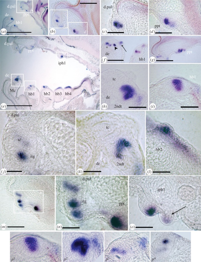
Ray-finned fishes (Actinopterygii) are the dominant vertebrate group today (+30 000 species, predominantly teleosts), with great morphological diversity, including their dentitions. How dental morphological variation evolved is best addressed by considering a range of taxa across actinopterygian phylogeny; here we examine the dentition of Polyodon spathula (American paddlefish), assigned to the basal group Acipenseriformes. Although teeth are present and functional in young individuals of Polyodon, they are completely absent in adults. Our current understanding of developmental genes operating in the dentition is primarily restricted to teleosts; we show that shh and bmp4, as highly conserved epithelial and mesenchymal genes for gnathostome tooth development, are similarly expressed at Polyodon tooth loci, thus extending this conserved developmental pattern within the Actinopterygii. These genes map spatio-temporal tooth initiation in Polyodon larvae and provide new data in both oral and pharyngeal tooth sites. Variation in cellular intensity of shh maps timing of tooth morphogenesis, revealing a second odontogenic wave as alternate sites within tooth rows, a dental pattern also present in more derived actinopterygians. Developmental timing for each tooth field in Polyodon follows a gradient, from rostral to caudal and ventral to dorsal, repeated during subsequent loss of teeth. The transitory Polyodon dentition is modified by cessation of tooth addition and loss. As such, Polyodon represents a basal actinopterygian model for the evolution of developmental novelty: initial conservation, followed by tooth loss, accommodating the adult trophic modification to filter-feeding.

Keywords: Polyodon; bmp4; dentition; evolution; paddlefish; shh.


Figures

Figure 1.
Figure 1.
Expression of shh, bmp4 in Polyodon spathula oral and pharyngeal initial dentitions, stage 41. (a–c,e) shh expression in tooth buds of cleared whole mount jaws compared with (d) stage 37 upper jaw, expression restricted to oral surfaces and on first infrapharygobranchial arches. (a,c) Multiple loci on tooth fields of dentary and dermopalatine, only two loci on hypobranchial and palatopterygoid. Arrows indicate alternate timing of strongest expression. (b,e) Strong expression in hypobranchial 1 and palatopterygoid (arrowheads); cone expression in dentary, hypobranchial, dermopalatine, compared to early placode expression on palatopterygoid. (f–i) bmp4 expression for comparison to shh expression. (f,g) Lower jaw, (h,i) upper jaw bmp4 in the dental papillary mesenchyme marks all oral jaw tooth positions. Dental mesenchyme underlies the dental epithelium and expression appears diffuse, however, more intense expression is seen at alternate tooth loci (arrows, f,g,i) with weaker expression indicating earlier (older) loci (asterisk), equivalent to shh expression pattern. Abbreviations: b1, 2, basibranchials; ba, bone of attachment, cb1–5, ceratobranchials; ch, ceratohyal; de, dentary; d.pal, dermopalatine; hb1, 2, 1st, 2nd hypobranchial; hb1tp, hb2tp, hypobranchial toothplates; hh, hypohyal; hym, hyomandibular; itg, incipient tooth germ; iph, infrapharyngobranchial; iphtp, infrapharyngobranchial toothplate; Mc, Meckels's cartilage; ppt, palatopterygoid; tc, tooth cone; 2ndt, second tooth.
Figure 2.
Figure 2.
Alizarin red, Alcian blue preparations of Polyodon spathula, 7dps showing relative tooth positions. (a, c, g-k) Upper jaw and dorsal pharyngeal skeleton, (b,d–f,l) lower jaw and ventral pharyngeal skeleton. (a,b) Chondrocranium and branchial arches. (c) Upper jaw, teeth along dermopalatine bone and separate palatopterygoid tooth plate (lacking membrane bone), with two paired tooth plates caudally (black arrows indicate j, k). (d) Lower jaw, ventral pharyngeal skeleton (hyoid, 1st, 2nd gill arches). (e) Teeth on dentary bone (arrows, new teeth). (f) Eight teeth linked by bone of attachment on 1st gill arch cartilage (hypobranchial 1, lacking membrane bone). (g–i) Right upper jaw, teeth ankylosed to dermopalatine bone, separate palatopterygoid tooth plate (arrows, new teeth caudally on dermopalatine (i), rostrally on palatopterygoid (h)). (h) Palatopterygoid tooth plate, bone of attachment only (arrows new teeth). (j,k) Upper jaw tooth plates of (j) epibranchial 2, four associated teeth, (k) hyoid arch, six teeth. (l) Hypohyal and first two ventral gill arches, with paired toothplates, more teeth on hb1 than hb2, more on ventral than dorsal pharyngeal toothplates. White arrows = newest unattached teeth. Scale bars (a,b), 1 mm; (c–g,l), 500 μm; (h,i), 100 μm; abbreviations as in figure 1.
Figure 3.
Figure 3.
Serial sagittal sections, Polyodon spathula (stage 45) after in situ hybridization for shh show sequence of tooth morphogenesis. Photomicrographs, low and high magnification (objectives 6.3×, 16×, 40×) of location and rostro-caudal timing of shh gene expression in all tooth fields relative to tooth germ morphogenesis, rostral, left and dorsal, top. (a–d) Most medial section, expression in dermopalatine (cone + collar, p3) and palatopterygoid (placode, p1). (e) More lateral section including Meckel's cartilage and pharyngeal arches. Expression loci associated with first stages of morphogenesis (placode, p1) on the 1st upper branchial arch (iph1), 1st and 2nd hypobranchials. By comparison, on 3rd and 4th pharyngeal arches tooth bud foci absent, localization is a field of expression, a stage prior to tooth morphogenesis. (f) Low magnification field of variation in expression loci on dentary and hypobranchial1, with collar epithelium downregulated on first tooth (asterisk) and adjacent second tooth germ shown as intense expression (arrowhead, weak expression in sensory papilla, arrow as (o, p4). (g) Low magnification view of variation in expression at loci on the dermopalatine (downregulated) and palatopterygoid strong expression in all dental epithelium around dentine cone (late cap stage). (h) Tooth cone (tc) developed, and 2nd tooth germ (2ndt) at cap stage (p2). (i) First hypobranchial, placode stage of shh expression (p1). (j) Tooth cone with second incipient tooth germ (itg), strong expression (p4). (k) Downregulation from cap to ‘collar’ expression (p3) in 2nd tooth. (l) Early tooth placode in oral epithelium of 2nd hypobranchial. (m) Upper jaw palatoquadrate cartilage with tooth germs on dermopalatine and palatopterygoid at different morphogenetic stages. (n) Four stages of shh expression, tooth cone with downregulated expression, incipient second tooth germ on dermoplatine, on palatopterygoid, cap stage. (o) Infrapharyngobranchial (iph1) upregulated strong expression (note evaginated tooth germ, placode-cap), alongside weak expression in sensory papilla (arrow). (p14) Four stages of shh expression in tooth germs, oral epithelium dorsal, contrast enhanced (translated into diagram as figure 4a–d). Scale bars (a,e), 250 μm; (b,f,g,m), 50 μm; (c,d,h–l,n–p14), 25 μm; abbreviations as in figure 1.
Figure 4.
Figure 4.
Diagram summarizing stages of tooth germ morphogenesis relative to shh expression (from figure 3p1–4). Intensity of cellular expression is partitioned characteristically within the dental epithelium, with negative differentiated, interactive cells of dental epithelium shown (proactive d.e.), and also in sensory papilla of taste buds on right of tooth germ. (a) Cellular partitioning of shh expression as ‘placode’ (localized within epithelium, can be evaginated). (b) ‘Cap’, expression in the cap-shaped epithelium of tooth germ, surrounding dental papilla. (c) ‘Cone + collar’, cones of dentine with expression associated with the tooth base, or collar epithelium below the cap. (d) ‘Cone + bud’, expression in a new site within the outer dental epithelia (incipient bud for new tooth germ).

References

    1. Grande L, Bemis WE. 1991. Osteology and phylogenetic relationships of fossil and recent paddlefishes (Polyodontidae) with comments on the interrelationships of Acipenseriformes. J. Vert. Paleo. Spec. Mem. 1, 1–121. (10.1080/02724634.1991.10011424) - DOI
    1. Bemis WE, Findeis EK, Grande L. 1997. An overview of Acipenseriformes. Environ. Biol. Fishes 48, 25–71. (10.1023/A:1007370213924) - DOI
    1. Bemis WE, Grande L. 1992. Early development of the actinopterygian head. I. External development and staging of the paddlefish Polyodon spathula . J. Morph. 213, 47–83. (10.1002/jmor.1052130106) - DOI - PubMed
    1. Davis MC. 2013. The deep homology of the autopod: insights from Hox gene regulation. Int. Comp. Biol. 53, 224–234. (10.1093/icb/ict029) - DOI - PubMed
    1. Modrell MS, Buckley D, Baker CVH. 2011. Molecular analysis of neurogenic placode development in a basal ray-finned fish. Genesis 49, 278–294. (10.1002/dvg.20707) - DOI - PMC - PubMed

Publication types

LinkOut - more resources