. 2015 Mar 18;35(11):4528-39.
doi: 10.1523/JNEUROSCI.1188-14.2015.

Regional and stage-specific effects of prospectively purified vascular cells on the adult V-SVZ neural stem cell lineage

Affiliations

Regional and stage-specific effects of prospectively purified vascular cells on the adult V-SVZ neural stem cell lineage

Elizabeth E Crouch et al. J Neurosci. .

Abstract

Adult neural stem cells reside in specialized niches. In the ventricular-subventricular zone (V-SVZ), quiescent neural stem cells (qNSCs) become activated (aNSCs), and generate transit amplifying cells (TACs), which give rise to neuroblasts that migrate to the olfactory bulb. The vasculature is an important component of the adult neural stem cell niche, but whether vascular cells in neurogenic areas are intrinsically different from those elsewhere in the brain is unknown. Moreover, the contribution of pericytes to the neural stem cell niche has not been defined. Here, we describe a rapid FACS purification strategy to simultaneously isolate primary endothelial cells and pericytes from brain microregions of nontransgenic mice using CD31 and CD13 as surface markers. We compared the effect of purified vascular cells from a neurogenic (V-SVZ) and non-neurogenic brain region (cortex) on the V-SVZ stem cell lineage in vitro. Endothelial and pericyte diffusible signals from both regions differentially promote the proliferation and neuronal differentiation of qNSCs, aNSCs, and TACs. Unexpectedly, diffusible cortical signals had the most potent effects on V-SVZ proliferation and neurogenesis, highlighting the intrinsic capacity of non-neurogenic vasculature to support stem cell behavior. Finally, we identify PlGF-2 as an endothelial-derived mitogen that promotes V-SVZ cell proliferation. This purification strategy provides a platform to define the functional and molecular contribution of vascular cells to stem cell niches and other brain regions under different physiological and pathological states.

Keywords: adult neurogenesis; endothelial cells; neural stem cell; niche; pericytes; vascular.


Figures

Figure 1.
Figure 1.
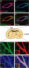
CD13 labels pericytes in the adult mouse cortex and V-SVZ. Center, Schema of coronal section of the adult mouse brain showing location of cortex (Ctx) and V-SVZ. Shown are confocal z-stack projections of immunostaining for CD13, CD31, and PDGFR-β in coronal sections of the cortex (AD) and whole-mount preparations of the V-SVZ (EH). Arrowheads in EH point to pericyte soma, which protrude off of blood vessels. Insets show higher magnifications of three merged optical slices from the z-stack in the boxed area in EH. Both CD13 and PDGFR-β label pericytes (B, D, E, F, H). Neither CD13 nor PDGFR-β colabel CD31+ endothelial cells (A, C, D, G, H). Scale bars, 10 μm; inset, 30 μm.
Figure 2.
Figure 2.
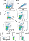
FACS isolation strategy for primary endothelial cells and pericytes from brain microregions. Representative FACS plots showing the gating strategy for the purification of endothelial cells and pericytes from the adult mouse brain. After excluding debris (A), doublets (B), and dead cells (DAPI+; C), CD45CD41 cells are selected (D) and CD31+CD13 endothelial cells and CD31CD13+ pericytes are collected (Ctx, E; V-SVZ, F). Images in DF are displayed using auto biexponential display. Percentages refer to the average proportion of cells in the previous parent gate. Isotype controls and the percentage of nonspecific labeling for each fluorophore are shown in G (FITC), H (PE), and I (APC).
Figure 3.
Figure 3.
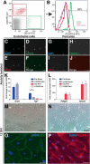
Characterization of FACS-purified endothelial cells and pericytes. A, B, By flow cytometry, >99% of CD31+CD13 endothelial cells (red gate, A) are PDGFR-β+ (B) and, conversely, CD31CD13+ pericytes (green gate, A) are PDGFR-β+ (B). Blood cells are shown in fuchsia in B. CF, Images of acutely immunostained purified endothelial cells. Purified endothelial cells (DAPI, C) are colabeled with CD105 (D) and CD31 (E). F is a merged image. GJ, Images of acutely immunostained purified pericytes. Purified pericytes (DAPI, G) are colabeled with PDGFR-β (H) and CD13 (I). J is a merged image. K, L, qPCR validation of expression of endothelial and pericyte genes in acutely purified populations. Expression levels of endothelial genes Glut1 and Tjp1 (K) and pericyte genes Pdgfrb and Abcc9 (L) compared with total V-SVZ. (n= >3, mean ± SEM; *p < 0.05, unpaired Student's t test). M, N, Phase images of endothelial cells (M) and pericytes (N) after 2 weeks in culture. O, P, Immunostaining for CD31 in cultured endothelial cells (O) and PDGFR-β in cultured pericytes (P). Scale bars: F, J, 100 μm; MP, 70 μm.
Figure 4.
Figure 4.
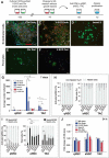
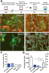
Effect of endothelial and pericyte-diffusible signals on V-SVZ stem cell proliferation. A, Schema of experimental paradigm to test the effect of diffusible signals from vascular cells on specific stages of the V-SVZ stem cell lineage. BF, Typical morphology of aNSC colonies cultured with conditioned medium (CM) from cortical (Ctx) endothelial cells (B), V-SVZ endothelial cells (C), the bend.3 endothelial cell line (D), cortical pericytes (E), and V-SVZ pericytes (F). G, Quantification of average number of cells per well generated by qNSCs, aNSCs, and TACs with different conditioned medium (mean ± SEM, n = 6, *p < 0.05; **p < 0.01, one-way ANOVA followed by post hoc Bonferroni's multiple-comparisons test. p-values for cortical endothelial cells versus all other conditions are p < 0.01 for aNSCs and p < 0.05 for TACs. H, Quantification of nestin and TuJ1 immunostaining in V-SVZ cell populations at 7 d in vitro after culture with different conditioned media. I, Quantification of Nestin and Ki67 (qNSCs, 7 d) and of Nestin and MCM2 (aNSCs and TACs, 4 d) immunostaining after exposure to different conditioned media. J, Quantification of the effect of conditioned media on survival of qNSCs, aNSCs, and TACs at 24 h in vitro (n = 3, mean ± SEM, *p < 0.05; **p < 0.01, one-way ANOVA followed by post hoc Bonferroni's multiple-comparisons test). Scale bar, 100 μm.
Figure 5.
Figure 5.
Primary endothelial cell signals increase the proliferation of existing aNSC clones. A, Schema outlining experimental paradigm to test the effect of diffusible signals from endothelial cells on the proliferation aNSCs in vitro at the single cell level. BE, Typical colonies generated by single aNSCs in response to conditioned medium (CM) from primary cortical (B) and V-SVZ (C) endothelial cells, the bend.3 cell line (D), or EGF alone (E). F, Quantification of activated clones. The difference between conditions is not significant by ANOVA (p = 0.788). G, Quantification of average size of single-cell clones (n = 3, mean ± SEM, *p < 0.05, one-way ANOVA followed by post hoc Bonferroni's multiple-comparisons test). Scale bars in BE, 200 μm.
Figure 6.
Figure 6.
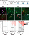
Effect of endothelial and pericyte-diffusible signals on differentiation. A, Schema outlining experimental paradigm to test the effect of diffusible signals in conditioned medium (CM) from endothelial cells and pericytes on the differentiation of qNSCs, aNSCs, and TACs in vitro. BC, Cortical pericyte signals produced two types of colonies from qNSCs: rare colonies with a single central astrocyte and chains of neurons (B) and colonies similar to those produced by aNSCs (C). DH, Image of aNSCs cultured with cortical (D), V-SVZ (E), or bend.3 (F) endothelial conditioned medium; cortical (G) or V-SVZ (H) pericyte-conditioned medium; or EGF (I). J, Quantification of differentiation (n = 3, mean ± SEM; *p < 0.05; **p < 0.01, one-way ANOVA followed by post hoc Bonferroni's multiple-comparisons test). Scale bars: B, 40 μm; CI, 50 μm.
Figure 7.
Figure 7.
Endothelial-derived PIGF-2 promotes V/SVZ proliferation. A, Antibody arrays of conditioned medium from cortical and V/SVZ endothelial cells and pericytes. Duplicate dots show the levels of VEGF-A, VEGF-B, PIGF-2, and SerpinE1. B, Relative pixel intensity of expression of VEGF-A, VEGF-B, PIGF-2, and SerpinE1 in conditioned medium from V-SVZ and cortical endothelial cells and pericytes relative to positive control. C, Quantification of total cell number of aNSCs cultured at the concentrations indicated of the VEGFR1 and 2 inhibitor ZM 306416 together with EGF, PIGF-2, cortical endothelial conditioned medium, or neurosphere medium alone (n = 3). At higher doses (1 μm), nonspecific effects were observed. D, Quantification of total cell number of qNSCs, aNSCs, and TACs cultured with PIGF-2 alone, EGF alone, or NS medium alone. E, Percentage of activated qNSCs as determined by MCM2/Nestin+ staining. Error bars represent SEM. *p < 0.05; **p < 0.01; ***p < 0.001; ns, not significant.
Figure 8.
Figure 8.
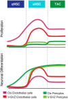
Model of the effect of diffusible signals from primary endothelial cells and pericytes on V-SVZ neural stem cell proliferation and neurogenesis. Top, Primary endothelial cells and pericytes from the V-SVZ and cortex (Ctx; non-neurogenic region) all increased the proliferation of aNSCs and TACs. Endothelial cell signals were more potent than their pericyte counterparts. Cortical endothelial cells alone were able to increase the proliferation of qNSCs. Bottom, Primary endothelial cells and pericytes from the V-SVZ and Ctx all increased neuronal differentiation of aNSCs and TACs. Only cortical pericytes increased neuronal differentiation from qNSCs.

References

    1. Andreu-Agulló C, Morante-Redolat JM, Delgado AC, Fariñas I. Vascular niche factor PEDF modulates Notch-dependent stemness in the adult subependymal zone. Nat Neurosci. 2009;12:1514–1523. doi: 10.1038/nn.2437. - DOI - PubMed
    1. Armulik A, Genové G, Mäe M, Nisancioglu MH, Wallgard E, Niaudet C, He L, Norlin J, Lindblom P, Strittmatter K, Johansson BR, Betsholtz C. Pericytes regulate the blood-brain barrier. Nature. 2010;468:557–561. doi: 10.1038/nature09522. - DOI - PubMed
    1. Armulik A, Genové G, Betsholtz C. Pericytes: developmental, physiological, and pathological perspectives, problems, and promises. Dev Cell. 2011;21:193–215. doi: 10.1016/j.devcel.2011.07.001. - DOI - PubMed
    1. Bardehle S, Krüger M, Buggenthin F, Schwausch J, Ninkovic J, Clevers H, Snippert HJ, Theis FJ, Meyer-Luehmann M, Bechmann I, Dimou L, Götz M. Live imaging of astrocyte responses to acute injury reveals selective juxtavascular proliferation. Nat Neurosci. 2013;16:580–586. doi: 10.1038/nn.3371. - DOI - PubMed
    1. Bell RD, Winkler EA, Sagare AP, Singh I, LaRue B, Deane R, Zlokovic BV. Pericytes control key neurovascular functions and neuronal phenotype in the adult brain and during brain aging. Neuron. 2010;68:409–427. doi: 10.1016/j.neuron.2010.09.043. - DOI - PMC - PubMed

Publication types