. 2015 Mar 18;35(11):4614-25.
doi: 10.1523/JNEUROSCI.1580-14.2015.

Nociceptor beta II, delta, and epsilon isoforms of PKC differentially mediate paclitaxel-induced spontaneous and evoked pain

Affiliations

Nociceptor beta II, delta, and epsilon isoforms of PKC differentially mediate paclitaxel-induced spontaneous and evoked pain

Ying He et al. J Neurosci. .

Abstract

As one of the most effective and frequently used chemotherapeutic agents, paclitaxel produces peripheral neuropathy (paclitaxel-induced peripheral neuropathy or PIPN) that negatively affects chemotherapy and persists after cancer therapy. The mechanisms underlying this dose-limiting side effect remain to be fully elucidated. This study aimed to investigate the role of nociceptor protein kinase C (PKC) isoforms in PIPN. Employing multiple complementary approaches, we have identified a subset of PKC isoforms, namely βII, δ, and ϵ, were activated by paclitaxel in the isolated primary afferent sensory neurons. Persistent activation of PKCβII, PKCδ, and PKCϵ was also observed in the dorsal root ganglion neurons after chronic treatment with paclitaxel in a mouse model of PIPN. Isoform-selective inhibitors of PKCβII, PKCδ, and PKCϵ given intrathecally dose-dependently attenuated paclitaxel-induced mechanical allodynia and heat hyperalgesia. Surprisingly, spinal inhibition of PKCβII and PKCδ, but not PKCϵ, blocked the spontaneous pain induced by paclitaxel. These data suggest that a subset of nociceptor PKC isoforms differentially contribute to spontaneous and evoked pain in PIPN, although it is not clear whether PKCϵ in other regions regulates spontaneous pain in PIPN. The findings can potentially offer new selective targets for pharmacological intervention of PIPN.

Keywords: cancer; chemotherapy; pain; protein kinase C; taxane.


Figures

Figure 1.
Figure 1.
A, Paclitaxel-induced plasma membrane translocation of PKC isoforms in DRG neurons. Among the PKC isoforms expressed in rat DRG neurons, PKCβII, PKCδ, and PKCϵ, but not other isoforms, showed significant plasma membrane translocation, indicative of their activation upon paclitaxel treatment (10 nm, 1 h). The fluorescent intensity of each PKC isoform across the cell (indicated by the arrow) is illustrated in the chart. B, Quantitative analysis of paclitaxel-treated DRG neurons with PKC translocation. The soma area of DRG neurons was measured to determine the percentage of sensory neurons within each size classification (small: <600 μm2, medium: 600–1200 μm2, or large: >1200 μm2; Peters et al., 2007) that had translocation of each PKC isoform (PKCβII, PKCδ, and PKCϵ). C, Size distribution of sensory neurons with PKC isoform (PKCβII, PKCδ, and PKCϵ) translocation by soma diameter. Six hundred neurons were examined for each isoform.
Figure 2.
Figure 2.
A, Translocation of PKC isoform (PKCβII, PKCδ, or PKCϵ) occurred in both IB4-positive and IB4-negative sensory neurons. Paclitaxel-treated DRG neurons were colabeled with PKC isoform PKCβII, PKCδ, or PKCϵ and IB4-FITC. B, Quantitative analysis of PKC translocation DRG neurons within IB4-positive and IB4-negative population (n = 300 neurons for each isoform). C, Translocation of PKCβII, PKCδ, or PKCϵ occurred in sensory neurons expressing TRPV1. Paclitaxel-treated DRG neurons were costained with PKC isoform (PKCβII, PKCδ, or PKCϵ) and TRPV1. Scale bar, 20 μm.
Figure 3.
Figure 3.
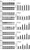
Paclitaxel (10 nm, 1 h) induced-PKCβII, PKCδ, and PKCϵ translocation from cytosol to plasma membrane as determined by cell fractionation followed by Western blotting analysis in the DRG neurons. *p < 0.05, **p < 0.01, ***p < 0.001 versus the cytosolic 0 time group; #p < 0.05, ##p < 0.01, ###p < 0.001 versus the membrane 0 time group (n = 3). The molecular weight markers are indicated by formula image(80 kDa) and formula image(42 kDa).
Figure 4.
Figure 4.
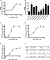
A, Dose-dependent release of CGRP produced by nanomolar concentrations of paclitaxel in the DRG neurons. B, Inhibition of PKCβII, PKCδ, and PKCϵ, but not other isoforms, significantly reduced the release of CGRP induced by paclitaxel. Individual myristoylated peptide inhibitor of PKC isoforms (10 μm) was preincubated with DRG cells for 10 min and present throughout the 10 min incubation for release assay. **p < 0.01, ***p < 0.001 versus the control group; ## p < 0.01 versus the paclitaxel group (n = 3). C–F, Dose–response curves and analysis of the inhibitors of PKCβII, PKCδ, and PKCϵ on the suppression of paclitaxel-induced CGRP release (n = 3). IC50 and Emax for each inhibitor were determined based on the dose–response curve. §§p < 0.01, §§§p < 0.001 versus the “βII” group; ¶¶p < 0.01 versus the “δ” group.
Figure 5.
Figure 5.
Activation of PKCβII, PKCδ, and PKCϵ in primary afferent neurons in a mouse model of PIPN. A, PIPN was induced by paclitaxel (1 mg/kg, i.p.; every other day for 4 treatments in ICR mice). Isoform-selective inhibitors of PKCβII (3.0 nmole), PKCδ (3.0 nmole), and PKCϵ (1.6 nmole) were administered intrathecally on day 26. B, Paclitaxel-induced plasma membrane translocation of PKCβII, PKCδ, and PKCϵ was present in DRG neurons, which was significantly abolished 30 min after spinal administration of the PKC isoform inhibitors, respectively (bottom). Scale bar, 20 μm.
Figure 6.
Figure 6.
Dose- and time-dependent attenuation of mechanical and thermal hypersensitivity by the inhibitors of PKCβII (A, B), PKCδ (C, D), and PKCϵ (E, F) in PIPN. The paw withdrawal threshold to von Frey filament probing (A, C, E) and withdrawal latency to radiant heat (B, D, F) were measured before (0) and 0.5, 1, 2, 4, and 8 h after the injection of PKC inhibitors (in 5 μl of saline, i.t.). *p < 0.05, **p < 0.01,***p < 0.001 versus the control group; #p < 0.05, ##p < 0.01, ###p < 0.001 versus the paclitaxel group; n = 8 for each group.
Figure 7.
Figure 7.
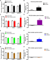
A, Lidocaine (0.04% in 5 μl of saline, i.t.) induced CPP in paclitaxel-treated mice. Paclitaxel mice spent significantly more time in the lidocaine-paired chamber, whereas control mice showed no chamber preference. B, Difference scores confirmed that paclitaxel-treated mice but not control mice showed CPP to lidocaine. C, E, Inhibitors of PKCβII (3.0 nmole in 5 μl of saline, i.t.) and PKCδ (3.0 nmole in 5 μl of saline, i.t.) produced CPP in paclitaxel mice. Paclitaxel-treated mice spent significantly more time in the inhibitor-paired chamber, whereas control mice showed no chamber preference. D, F, Difference scores confirmed that paclitaxel-treated mice, but not control mice, showed CPP to inhibitors of PKCβII and PKCδ. G, PKCϵ inhibitor (1.6 nmole in 5 μl of saline, i.t.) did not produce CPP in paclitaxel or control mice. Paclitaxel-treated mice and control mice showed no chamber preference, spending similar amount of time in saline- and PKCϵ inhibitor-paired chambers. H, Difference scores confirmed the absence of chamber preference. *p < 0.05; **p < 0.01; ***p < 0.001; n = 8 for each group.

Comment in

References

    1. Alessandri-Haber N, Dina OA, Chen X, Levine JD. TRPC1 and TRPC6 channels cooperate with TRPV4 to mediate mechanical hyperalgesia and nociceptor sensitization. J Neurosci. 2009;29:6217–6228. doi: 10.1523/JNEUROSCI.0893-09.2009. - DOI - PMC - PubMed
    1. Amadesi S, Cottrell GS, Divino L, Chapman K, Grady EF, Bautista F, Karanjia R, Barajas-Lopez C, Vanner S, Vergnolle N, Bunnett NW. Protease-activated receptor 2 sensitizes TRPV1 by protein kinase Cepsilon- and A-dependent mechanisms in rats and mice. J Physiol. 2006;575:555–571. doi: 10.1113/jphysiol.2006.111534. - DOI - PMC - PubMed
    1. Barber LA, Vasko MR. Activation of protein kinase C augments peptide release from rat sensory neurons. J Neurochem. 1996;67:72–80. - PubMed
    1. Borsook D, Hargreaves R, Bountra C, Porreca F. Lost but making progress–where will new analgesic drugs come from? Sci Transl Med. 2014;6:249sr3. doi: 10.1126/scitranslmed.3008320. - DOI - PMC - PubMed
    1. Brager DH, Cai X, Thompson SM. Activity-dependent activation of presynaptic protein kinase C mediates post-tetanic potentiation. Nat Neurosci. 2003;6:551–552. doi: 10.1038/nn1067. - DOI - PubMed

MeSH terms