. 2015 Mar 18;35(11):4729-40.
doi: 10.1523/JNEUROSCI.3304-13.2015.

Secreted frizzled related proteins modulate pathfinding and fasciculation of mouse retina ganglion cell axons by direct and indirect mechanisms

Affiliations

Secreted frizzled related proteins modulate pathfinding and fasciculation of mouse retina ganglion cell axons by direct and indirect mechanisms

Séverine Marcos et al. J Neurosci. .

Abstract

Retina ganglion cell (RGC) axons grow along a stereotyped pathway undergoing coordinated rounds of fasciculation and defasciculation, which are critical to establishing proper eye-brain connections. How this coordination is achieved is poorly understood, but shedding of guidance cues by metalloproteinases is emerging as a relevant mechanism. Secreted Frizzled Related Proteins (Sfrps) are multifunctional proteins, which, among others, reorient RGC growth cones by regulating intracellular second messengers, and interact with Tolloid and ADAM metalloproteinases, thereby repressing their activity. Here, we show that the combination of these two functions well explain the axon guidance phenotype observed in Sfrp1 and Sfrp2 single and compound mouse mutant embryos, in which RGC axons make subtle but significant mistakes during their intraretinal growth and inappropriately defasciculate along their pathway. The distribution of Sfrp1 and Sfrp2 in the eye is consistent with the idea that Sfrp1/2 normally constrain axon growth into the fiber layer and the optic disc. Disheveled axon growth instead seems linked to Sfrp-mediated modulation of metalloproteinase activity. Indeed, retinal explants from embryos with different Sfrp-null alleles or explants overexpressing ADAM10 extend axons with a disheveled appearance, which is reverted by the addition of Sfrp1 or an ADAM10-specific inhibitor. This mode of growth is associated with an abnormal proteolytic processing of L1 and N-cadherin, two ADAM10 substrates previously implicated in axon guidance. We thus propose that Sfrps contribute to coordinate visual axon growth with a dual mechanism: by directly signaling at the growth cone and by regulating the processing of other relevant cues.

Keywords: Sfrp; axon guidance; metalloproteinase; optic chiasm; optic disc; visual pathway.


Figures

Figure 1.
Figure 1.
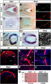
Expression of Sfrp1 and Sfrp2 in the proximal visual pathway. Transverse sections of E14.5 (A, B, G) and E13.5 (F, H, I-L′) retinas and E15.5 diencephalon (C, D) from wild-type (AD, FK′) and Sfrp2−/− (J, L, L′) embryos hybridized with probes for Sfrp1 (A, C) and Sfrp2 (F, G) or immunostained with α-Sfrp1 (B, D) or α-Sfrp2 antibodies (HL′). E, Dissociated culture from wt E14.5 retinas coimmunostained with α-Sfrp1 (green) and α-βIII-tubulin (red) antibodies. Note the expression of Sfrp1 in the RGC layer (RGC-l), retina pigmented epithelium (rpe), ciliary margins (cm), and lens (le). Note also that Sfrp1 mRNA is not present in the optic chiasm (och). Sfrp1 protein is abundantly detected in the RGC axons as they extend along the fiber layer (fl), optic nerve (on), and chiasm (B, D). This localization is confirmed in cultured neurons, where Sfrp1 localizes to the main neurite but it is absent from collateral branches (E, arrows). Sfrp2 mRNA localizes instead to the proliferating neural retina (nr), and the initial portion of the developing optic nerve (on, arrow in F) and the glial cells of the OD (od; arrowheads in G) and lens, but it is absent from differentiated RGCs and the ciliary margins (cmz). At E14.5, Sfrp2 expression in the dorsal-central retina begins to be downregulated (bracket in G). As demonstrated by the lack of any specific signal in Sfrp2 mutant embryos (J, L, L′), Sfrp2 protein was specifically detected not only in the retinal progenitors but also in the nascent retinal ganglion cells (rgc) and their axons (H, arrows) as well as in cells of the optic nerve (K′). Asterisks in IL indicate autofluorescent signal from the retina pigmented epithelium (rpe) and the red blood cells. M, Western blot analysis of Sfrp2 expression in heparin purified extracts of eyes from E14 wt and Sfrp2−/− embryos. Note the presence of a specific band in wt but not mutant retinas (right). Each lane contains an equal amount of proteins as defined by Ponceau staining (left). Scale bars: 250 μm (F, G); 200 μm (AD); 100 μm (IL); 20 μm (K′, L′).
Figure 2.
Figure 2.
Sfrp1/2 are required for correct intraretinal organization of RGC axons. Coronal sections (AC) and flat mounts (DI) of retinas from E15.5 wt, Sfrp2−/−, and Sfrp1−/−;Sfrp2−/− embryos immunostained with α-NF-M antibodies. Images in DI are confocal planes of the fiber (DF) and outer (GI) layers of the retinas. Note that, in wt, all RGC axons extend toward the fiber layer (fl, in A), whereas in the mutants, several axons from peripheral RGCs invade the outer retina (arrows in insets in B, C). Image in B shows the dorsal retina; images in A and C, the ventral retina. Axons invading the outer retina are also observed in flat-mount preparations (H, I). Note also that, in compound mutants, RGC axon bundles in the fiber layer are less defined and often present axons abnormally extending away from the OD (od, arrowheads in F). J, Mean ± SEM number of ectopic axons per coronal section in the dorsal or ventral periphery of wt (n = 6), Sfrp1−/− (n = 10), Sfrp2−/− (n = 6), and Sfrp1−/−;Sfrp2−/− (n = 6) deficient retinas. *p < 0.05 compared with wt. K, Mean ± SEM number of axons or small axon bundles located in the OD region, in the dorsal or ventral outer retina in wt (n = 12), Sfrp1−/− (n = 19), Sfrp2−/− (n = 14), and Sfrp1−/−;Sfrp2−/− (n = 8) flat-mounted retinal preparations. *p < 0.05; **p < 0.01; ***p < 0.001 compared with wt; Student's t test. LN, Coronal sections of wt (L), Sfrp2−/− (M), and Sfrp1−/−;Sfrp2−/− (N) E15.5 retinas after DiI retrograde tracings from the optic tract. Note that, in Sfrp2−/− and Sfrp1−/−;Sfrp2−/− embryos, few traced axons take abnormal trajectories. Please note the increased number of RGCs in the double mutant (asterisk in N) as already described (Esteve et al., 2011b). Scale bars, 200 μm (AI; LN).
Figure 3.
Figure 3.
A proportion of RGC axons fail to enter the OD in the absence of Sfrp2. A, Schematic representation of the tracing strategy using crystals of DiI (red) and DiO (green) in the dorsal (d) and ventral (v) retina, respectively. BG, Fluorescent images of flat-mounted preparations from E15.5 wt (B), Sfrp1−/− (C), Sfrp2−/− (D), and Sfrp1−/−;Sfrp2−/− (EG) retinas. Dashed lines indicate the position of the OD. Note that wt axons from the dorsal and ventral retina extend straight into the OD (B), whereas a small proportion of axons avoid the OD in mutants (arrows, CG). H, Percentage of DiI- and DiO-labeled retinas with axons that fail to enter or detour around the OD in wt (n = 14), Sfrp1−/− (n = 36), Sfrp2−/− (n = 20), and Sfrp1−/−;Sfrp2−/− (n = 30). Scale bars, 200 μm (BG).
Figure 4.
Figure 4.
Sfrp's are required for proper growth of RGC axons at the chiasm. AF, Ventral views of intact brain preparations from E15.5 wt (A), Sfrp1−/− (B), Sfrp2−/− (C), and Sfrp1−/−;Sfrp2−/− (DF) embryos. Axons from the right eye were traced by the insertion of DiI crystals into the OD. Anterior is to the top, posterior to the bottom. Arrowheads indicate the midline. Insets in AD are high magnifications with inverted contrast of the prechiasmatic end of the optic nerve. In wt and Sfrp1−/− (A, B), RGC axons extend into the optic nerve (ON) in tight bundles and slightly defasciculate as they enter the optic chiasm (inset). The majority of the axons enter the contralateral optic tract (cOT), whereas a small proportion remain uncrossed and grow into the ipsilateral optic tract (iOT) or enter the contralateral optic nerve (cON). The prechiasmatic end of the ON appears more defasciculated in Sfrp2−/− and Sfrp1−/−;Sfrp2−/− (arrows and insets in CE). Note also the presence of small axon bundles (arrowhead in E). F, In extreme cases, fibers appear completely defasciculated, equally divided into the ipsilateral and contralateral tracts, with many axons entering the contralateral optic nerve. G, H, Mean ± SEM of the prechiasmatic dispersion of the optic nerve at E13.5 and E15.5. **p < 0.01; ***p < 0.001 compared with wt (Student's t test). Scale bars, 200 μm (AF).
Figure 5.
Figure 5.
Sfrp1 and Sfrp2 deficiency causes premature defasciculation of axons in the optic tract. AH, Lateral views of DiI-labeled RGC axons as they grow along the contralateral optic tract in whole brain preparations from E15.5 wt (A, E), Sfrp1−/− (B, F), Sfrp2−/− (C, G), and Sfrp1−/−; Sfrp2−/− (D, H) embryos. The tract was exposed after cortex removal. In all images, dorsal is up and caudal is to the right. Open arrows in AD point to the level in which the optic tract starts to fan out, which is slightly distinct among the genotypes. EH, Images are higher magnifications at the level of the lateral geniculate nucleus (LGN) of those shown in AD. The contrast has been inverted to better appreciate the caudal dispersion of labeled fibers (arrowheads). Double arrowed lines in EH indicate the width occupied by DiI-labeled fibers as they approach the LGN. I, Mean ± SEM of the optic tract width in wt (n = 11), Sfrp1−/− (n = 11), Sfrp2−/− (n = 10), and Sfrp1−/−;Sfrp2−/− (n = 11) embryos at the level indicated in EH. J, Percentage of DiI-labeled wt and mutant embryos with an evident caudal spreading of the optic tract. *p < 0.05; ***p < 0.001 compared with wt (Student's t test). K, L, Fluorescent images with inverted contrast of frontal sections at the level of the optic tract from wt (K) and Sfrp1−/−;Sfrp2−/− (L) E15.5 embryos after unilateral DiI filling of the visual fibers. Note the lateral to medial dispersion of the fibers in the compound mutants (arrow). Di, Diencephalon; OC, optic chiasm; SC, superior colliculus; Tel, telencephalon. Scale bars: 150 μm (AD), 100 μm (EH), 200 μm (K, L).
Figure 6.
Figure 6.
Fasciculated growth of RGC neurites requires modulation of ADAM activity by Sfrp's. AC, Transverse sections of E15.5 retinas from wt, Sfrp2−/− and Sfrp1−/−;Sfrp2−/− embryos hybridized with a probe against Adam10. Note the strong Adam10 expression in the RGC layer. In double mutants, the number of RGCs is increased as described previously (Esteve et al., 2011a). DO, Low- and high-power views of retinal explants from E15.5 wt (D, G, J, M), Sfrp2−/− (E, H, K, N) and Sfrp1−/−;Sfrp2−/− (F, I, L, O) embryos cultured alone (DI) or in the presence of Sfrp1 protein (JL) or GI254023X, a specific ADAM10 inhibitor (MO). P, Mean ± SEM width of neurite fascicles of retinal explants from wt or Sfrp1−/−;Sfrp2−/− embryos cultured in different conditions. Q, Graph representing the proportion of fascicles wider than 60 pixels (pxs) in wt and Sfrp1−/−;Sfrp2−/− retinal explants grown with or without the addition of Sfrp1. R, Mean ± SEM width of neurite fascicles of retinal explants from wt or Sfrp2−/− retinas grown with DMSO (control) or GI254023X. S, Graph representing the proportion of fascicles of given width from wt and Sfrp2−/− retinal explants grown with DMSO or GI254023X. *p < 0.05; ***p < 0.001, Student's t test. Scale bars: 200 μm (AC), 200 μm (DF), 10 μm (GO).
Figure 7.
Figure 7.
ADAM10 mediates disheveled RGC axon growth likely by regulating L1 and N-cadherin proteolysis. AD, Transverse sections of E15.5 wt (A, B) and Sfrp1−/−;Sfrp2−/− (C, D) embryos at the level of the eye (A, C) and optic chiasm (B, D). Sections are immunostained with α-N-cadherin (A, C) and α-L1 (B, D) antibodies. E, F, Western blot analysis of L1 (E) and N-cadherin (F) processing in lysates of the proximal visual pathway from E16.5 wt, Sfrp2−/−, and Sfrp1−/−;Sfrp2−/− embryos. The 32 kDa L1 and 35 kDa N-cadherin fragments are increased in Sfrp2−/− and Sfrp1−/−;Sfrp2−/− embryos, as shown in the graph bar representation of processed and unprocessed bands normalized to α-tubulin as a loading control. The data represent a typical experiment. The lane showing the wt lysate derives from the same gel, in which an intermediate lane was eliminated. G, H, Confocal images of the retinal explants electroporated with GFP (G) or GFP+Adam10 (H)-immunostained with antibodies against GFP and β-III-tubulin. Note that ADAM10 overexpression causes axon defasciculation. I, Western blot analysis of GFP demonstrating similar levels of explant targeting. GFP levels were normalized against α-tubulin. J, Graph representing the proportion of fascicles of given width from retinal explants electroporated with GFP or Adam10. ADAM10 significantly reduces the number of wider fascicles. K, L, Western blot analysis of L1 (K) and N-cadherin (L) processing in lysates of GFP and Adam10-electroporated retinal explants. Note the increased ratio of processed/unprocessed N-cadherin and L1 normalized to α-tubulin in Adam10-overexpressing explants. on, Optic nerve; och (optic chiasm). *p < 0.05, ***p < 0.001; Bonferroni's test. Scale bars: 200 μm (AD), 10 μm (GH).

References

    1. Bai G, Pfaff SL. Protease regulation: the Yin and Yang of neural development and disease. Neuron. 2011;72:9–21. doi: 10.1016/j.neuron.2011.09.012. - DOI - PMC - PubMed
    1. Bao ZZ. Intraretinal projection of retinal ganglion cell axons as a model system for studying axon navigation. Brain Res. 2008;1192:165–177. doi: 10.1016/j.brainres.2007.01.116. - DOI - PMC - PubMed
    1. Bastmeyer M, Ott H, Leppert CA, Stuermer CA. Fish E587 glycoprotein, a member of the L1 family of cell adhesion molecules, participates in axonal fasciculation and the age-related order of ganglion cell axons in the goldfish retina. J Cell Biol. 1995;130:969–976. doi: 10.1083/jcb.130.4.969. - DOI - PMC - PubMed
    1. Birgbauer E, Cowan CA, Sretavan DW, Henkemeyer M. Kinase independent function of EphB receptors in retinal axon pathfinding to the optic disc from dorsal but not ventral retina. Development. 2000;127:1231–1241. - PubMed
    1. Blackshaw S, Harpavat S, Trimarchi J, Cai L, Huang H, Kuo WP, Weber G, Lee K, Fraioli RE, Cho SH, Yung R, Asch E, Ohno-Machado L, Wong WH, Cepko CL. Genomic analysis of mouse retinal development. PLoS Biol. 2004;2:E247. doi: 10.1371/journal.pbio.0020247. - DOI - PMC - PubMed

Publication types