

doi: 10.3892/ol.2015.2895.
Epub 2015 Jan 26.
Osteoid osteoma of the rib: A report of two cases
Affiliations
- PMID: 25789056
- PMCID: PMC4356267
- DOI: 10.3892/ol.2015.2895
Item in Clipboard
Osteoid osteoma of the rib: A report of two cases
Oncol Lett.
2015 Apr.
Abstract
Osteoid osteoma is type of benign bone tumor, characterized by a well-demarcated core with a typical size of <1 cm and by a distinctive surrounding zone of reactive bone formation. The tumor can occur anywhere in the cortex or medulla of the skeleton. However, the lesion usually affects the long bones of the lower extremities. The present study describes two cases of osteoid osteomas located in the rib.
Keywords: osteoid osteoma; rib; tumor.
Figures

X-ray image at anteroposterior view revealing the presence of a lesion at the tenth right rib.

(A) Computed tomography revealing the nidus and central calcification. (B) Three dimensional reconstructed view revealing a lesion located at the visceral side of the rib.

Bone scintigraphy revealing intense uptake at the site of the lesion.

Lesion was located during surgery using a C-arm fluoroscopic device.

(A) Specimen with the lesion at the visceral side. (B) The cross-section view revealing the location of the lesion in the cortex.

Photomicrograph revealing osteoid tissue in a background stroma of fibrovascular tissue (stain, hematoxylin and eosin; magnification, ×100).

Computed tomography revealing the location of the lesion at the top of sixth right rib.

Computed tomography revealing the location of the lesion at the top of sixth right rib.

X ray revealing the presence of scoliosis in the spine plain film.
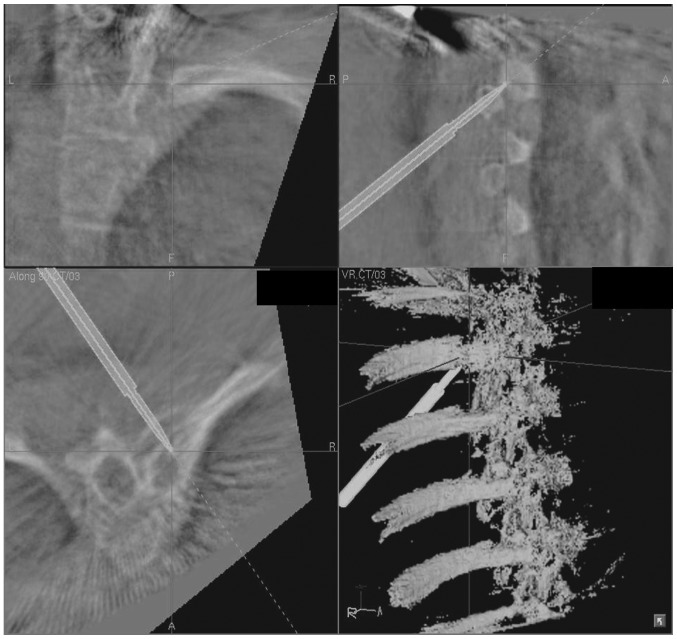
Intra-operative three dimensional C-arm-based navigation system was used to locate the tumor.

Photomicrograph revealing interconnecting trabeculae of woven bone lined prominently by osteoblasts (stain, hematoxylin and eosin; magnification, ×200).
Similar articles
-
Osteoid osteoma of the rib: A report of an extremely rare condition.Int J Surg Case Rep. 2022 May;94:107139. doi: 10.1016/j.ijscr.2022.107139. Epub 2022 May 6. Int J Surg Case Rep. 2022. PMID: 35658306 Free PMC article.
-
Osteoid osteoma of the rib masquerading as pain due to trauma: Removed by rib resection using preoperative CT-scan guidance.Int J Surg Case Rep. 2023 Feb;103:107877. doi: 10.1016/j.ijscr.2023.107877. Epub 2023 Jan 9. Int J Surg Case Rep. 2023. PMID: 36706671 Free PMC article.
-
Osteoid Osteoma.2023 Aug 14. In: StatPearls [Internet]. Treasure Island (FL): StatPearls Publishing; 2025 Jan–. 2023 Aug 14. In: StatPearls [Internet]. Treasure Island (FL): StatPearls Publishing; 2025 Jan–. PMID: 30725964 Free Books & Documents.
-
Osteoid osteoma: the uniquely innervated bone tumor.Mod Pathol. 1998 Feb;11(2):175-80. Mod Pathol. 1998. PMID: 9504688 Review.
-
Osteoid osteoma: multimodality imaging with focus on hybrid imaging.Eur J Nucl Med Mol Imaging. 2019 Apr;46(4):1019-1036. doi: 10.1007/s00259-018-4181-2. Epub 2018 Oct 19. Eur J Nucl Med Mol Imaging. 2019. PMID: 30341641 Review.
Cited by
-
Costal Osteoid Osteoma: A Case Report and Review of the Literature.Cureus. 2025 Jun 9;17(6):e85589. doi: 10.7759/cureus.85589. eCollection 2025 Jun. Cureus. 2025. PMID: 40636595 Free PMC article.
-
Osteoid osteoma of the rib: A report of an extremely rare condition.Int J Surg Case Rep. 2022 May;94:107139. doi: 10.1016/j.ijscr.2022.107139. Epub 2022 May 6. Int J Surg Case Rep. 2022. PMID: 35658306 Free PMC article.
-
A Rare Case of an Osteoid Osteoma of the Rib Treated under Computed Tomography Guidance: A Case Report and Review of the Literature.Case Rep Oncol. 2015 Nov 21;8(3):509-14. doi: 10.1159/000441835. eCollection 2015 Sep-Dec. Case Rep Oncol. 2015. PMID: 26668573 Free PMC article.
-
CT-guided percutaneous cryoablation of an osteoid osteoma of the rib☆.Radiol Case Rep. 2019 Jan 4;14(3):400-404. doi: 10.1016/j.radcr.2018.12.010. eCollection 2019 Mar. Radiol Case Rep. 2019. PMID: 30627298 Free PMC article.
-
Osteoid osteoma of the rib masquerading as pain due to trauma: Removed by rib resection using preoperative CT-scan guidance.Int J Surg Case Rep. 2023 Feb;103:107877. doi: 10.1016/j.ijscr.2023.107877. Epub 2023 Jan 9. Int J Surg Case Rep. 2023. PMID: 36706671 Free PMC article.
References
-
- Jaffe HL. Osteoid osteoma. A benign osteoblastic tumorcomposed of osteoid and atypical bone. Arch Surg. 1935;31:709–728. doi: 10.1001/archsurg.1935.01180170034003. - DOI
-
- Unni KK, editor. Dahlin’s bone tumors: general aspects and data on 11,087 cases. 5th edition. Lippincott-Raven; Philadelphia, PA: 1996. Osteoid osteoma; pp. 121–130.
-
- Ghanem I, Collet LM, Kharrat K, et al. Percutaneous radiofrequency coagulation of osteoid osteoma in children and adolescents. J Pediatr Orthop B. 2003;12:244–252. - PubMed
-
- Atesok KI, Alman BA, Schemitsch EH, et al. Osteoid osteoma and osteoblastoma. J Am Acad Orthop Surg. 2011;19:678–689. - PubMed
LinkOut - more resources
Full Text Sources
Other Literature Sources