

Behaviour of endothelial cells in a tridimensional in vitro environment
- PMID: 25789323
- PMCID: PMC4350961
- DOI: 10.1155/2015/630461
Behaviour of endothelial cells in a tridimensional in vitro environment
Abstract
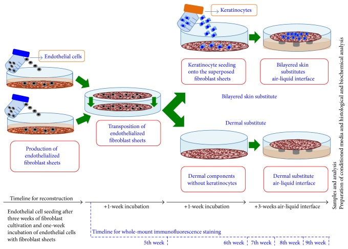
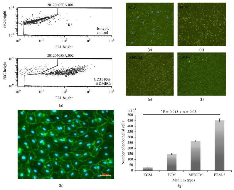
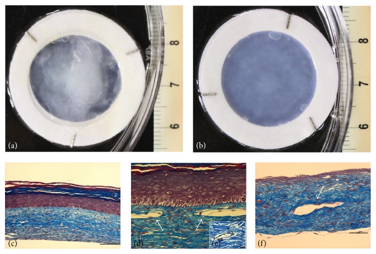
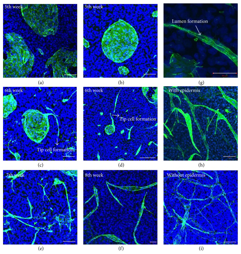
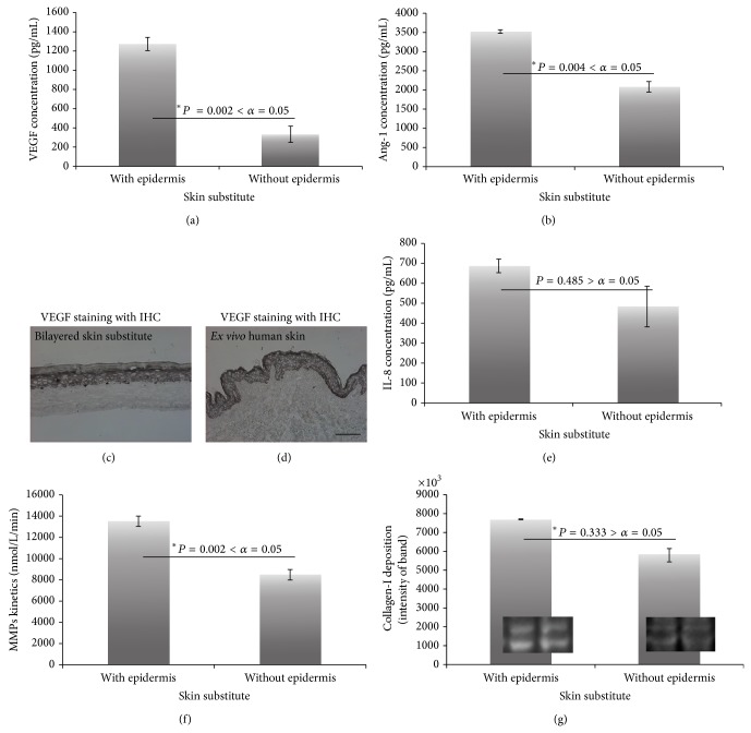
Angiogenesis is a fundamental process in healing, tumor growth, and a variety of medical conditions. For this reason, in vitro angiogenesis is an area of interest for researchers. Additionally, in vitro angiogenesis is important for the survival of prevascularized tissue-engineering models. The aim of this study was to observe the self-tubular organization behaviour of endothelial cells in the self-assembly method. In this study, bilayered and dermal substitutes were prepared using the self-assembly method. Histological, immunostaining, and biochemical tests were performed. The behavioural dynamics of endothelial cells in this biological environment of supportive cells were observed, as were the steps of the in vitro angiogenic cascade with self-organizing capillary-like structures formation. The epidermal component of the substitutes was seen to promote network expansion and density. It also increased the quantity of angiogenic factors (VEGF and Ang-1) without increasing the proinflammatory factor (IL-8). In addition, the increased MMP activity contributed to matrix degradation, which facilitated capillary formation.
Figures
Similar articles
-
Effect of living cellular sheets on the angiogenic potential of human microvascular endothelial cells.J Periodontol. 2015 May;86(5):703-12. doi: 10.1902/jop.2015.140362. Epub 2015 Jan 16. J Periodontol. 2015. PMID: 25594425
-
Vascular endothelial growth factor-C promotes vasculogenesis, angiogenesis, and collagen constriction in three-dimensional collagen gels.J Vasc Surg. 2005 Apr;41(4):699-707. doi: 10.1016/j.jvs.2005.01.015. J Vasc Surg. 2005. PMID: 15874936
-
A mathematical model for the capillary endothelial cell-extracellular matrix interactions in wound-healing angiogenesis.IMA J Math Appl Med Biol. 1997 Dec;14(4):261-81. IMA J Math Appl Med Biol. 1997. PMID: 9415995
-
Mechanisms of ocular angiogenesis and its molecular mediators.Dev Ophthalmol. 2010;46:4-20. doi: 10.1159/000320006. Epub 2010 Aug 10. Dev Ophthalmol. 2010. PMID: 20703029 Review.
-
In vitro models of angiogenesis.World J Surg. 2007 Apr;31(4):654-63. doi: 10.1007/s00268-006-0763-4. World J Surg. 2007. PMID: 17372665 Review.
Cited by
-
Biological activity of a vascular endothelial cell-hydroxyapatite orbital implant complex: An experimental study.Exp Ther Med. 2022 Mar;23(3):227. doi: 10.3892/etm.2022.11152. Epub 2022 Jan 18. Exp Ther Med. 2022. PMID: 35222704 Free PMC article.
-
In vitro psoriasis models with focus on reconstructed skin models as promising tools in psoriasis research.Exp Biol Med (Maywood). 2017 Jun;242(11):1158-1169. doi: 10.1177/1535370217710637. Exp Biol Med (Maywood). 2017. PMID: 28585891 Free PMC article.
-
Zebrafish as an Emerging Model Organism to Study Angiogenesis in Development and Regeneration.Front Physiol. 2016 Mar 8;7:56. doi: 10.3389/fphys.2016.00056. eCollection 2016. Front Physiol. 2016. PMID: 27014075 Free PMC article. Review.
-
Conditioned medium produced by fibroblasts cultured in low oxygen pressure allows the formation of highly structured capillary-like networks in fibrin gels.Sci Rep. 2020 Jun 9;10(1):9291. doi: 10.1038/s41598-020-66145-z. Sci Rep. 2020. PMID: 32518266 Free PMC article.
References
-
- Halama T., Staffler G., Hoch S., Stockinger H., Wolff K., Petzelbauer P. Vascular-endothelial cadherin (CD144)- but not PECAM-1 (CD31)-based cell-to-cell contacts convey the maintenance of a quiescent endothelial monolayer. International Archives of Allergy and Immunology. 1999;120(3):237–244. doi: 10.1159/000024273. - DOI - PubMed
-
- Lefleur M. A., Handsley M. M., Knäuper V., Murphy G., Edwards D. R. Endothelial tubulogenesis within fibrin gels specifically requires the activity of membrane-type-matrix metalloproteinases (MT-MMPs) Journal of Cell Science. 2002;115(17):3427–3438. - PubMed
Publication types
MeSH terms
Substances
LinkOut - more resources
Full Text Sources
Other Literature Sources
Miscellaneous
