. 2015:2015:494578.
doi: 10.1155/2015/494578. Epub 2015 Feb 19.

Alloxan-induced diabetes causes morphological and ultrastructural changes in rat liver that resemble the natural history of chronic fatty liver disease in humans

Affiliations

Alloxan-induced diabetes causes morphological and ultrastructural changes in rat liver that resemble the natural history of chronic fatty liver disease in humans

Amanda Natália Lucchesi et al. J Diabetes Res. 2015.

Abstract

Purpose: This study evaluated the long-term effects of alloxan-induced diabetes in rat liver.

Methods: Thirty nondiabetic control rats (NC) and 30 untreated diabetic (UD) rats were divided into three subgroups sacrificed after 6, 14, or 26 weeks. Clinical and laboratory parameters were assessed. Fresh liver weight and its relationship with body weight were obtained, and liver tissue was analyzed.

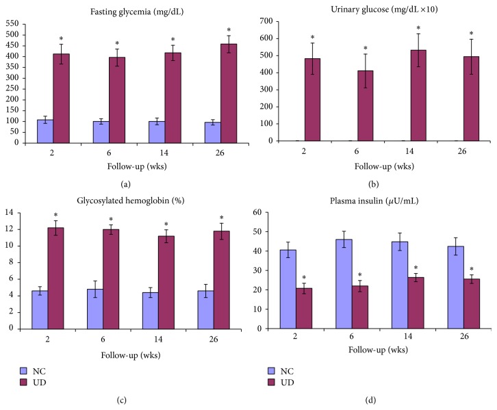
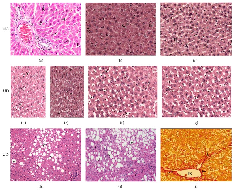
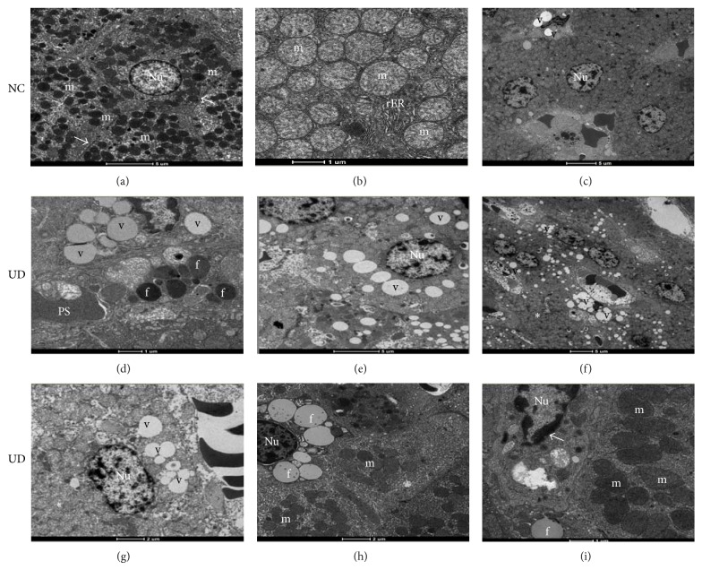
Results: UD rats showed sustained hyperglycemia, high glycosylated hemoglobin, and low plasma insulin. High serum levels of AST and ALT were observed in UD rats after 2 weeks, but only ALT remained elevated throughout the experiment. Fresh liver weight was equal between NC and UD rats, but the fresh liver weight/body weight ratio was significantly higher in UD rats after 14 and 26 weeks. UD rats showed liver morphological changes characterized by hepatic sinusoidal enlargement and micro- and macrovesicular hepatocyte fatty degeneration with progressive liver structure loss, steatohepatitis, and periportal fibrosis. Ultrastructural changes of hepatocytes, such as a decrease in the number of intracytoplasmic organelles and degeneration of mitochondria, rough endoplasmic reticulum, and nuclei, were also observed.

Conclusion: Alloxan-induced diabetes triggered liver morphological and ultrastructural changes that closely resembled human disease, ranging from steatosis to steatohepatitis and liver fibrosis.


Figures

Figure 1
Figure 1
Mean ± SD of fasting glycemia (a), urinary glucose (b), glycosylated hemoglobin (c), and plasma insulin (d) in nondiabetic control (NC) and untreated diabetic animals (UD) during follow-up time, given in weeks.
Figure 2
Figure 2
(a), (b), and (c): histological pattern of liver from nondiabetic control rats (NC) showing normal-appearing of hepatocytes, portal space (PS), sinusoids (arrows), and Kuppfer cells (K) at 6, 14, and 26 weeks of follow-up, respectively. Focal area of mild steatosis (∗) is observed in NC rats sacrificed at 26 weeks. (d) and (e): liver from untreated diabetic rats (UD) sacrificed at 6 weeks showing the onset of sinusoidal enlargement (arrows) and small amount of fatty vacuoles (v), respectively. (f) and (g): liver from UD rats sacrificed at 14 and 26 weeks showing progressive worsening of sinusoidal enlargement (arrows) and liver fatty degeneration (v), respectively. Liver from UD rats at 26 weeks showing in (h) macrovesicular fatty degeneration (v); (i) interlobular mononuclear inflammatory infiltrate consistent with steatohepatitis (∗); (j) periportal (PS) fibrosis (arrows). H&E and red picrosirius (400x).
Figure 3
Figure 3
Electron micrographs of nondiabetic control rats (NC) sacrificed at 6, 14, and 26 weeks, respectively, showing (a) normal-appearing of hepatocytes, nucleus (Nu), mitochondria (m) and Disse's space (arrows); (b) detail of mitochondria (m) and rough endoplasmic reticulum (rER) with their preserved structures; (c) focal area of lipid vacuoles (v) in NC rat sacrificed at 26 weeks (scale bars: (a), (c): 1,900x; (b): 4,800x). (d), (e), and (f): hepatocytes of untreated diabetic rats (UD) sacrificed at 6, 14, and 26 weeks, respectively, showing (d) hepatocyte showing moderate amount of lipid vacuoles (v) and hepatic lipid droplets (f); (e) worsening of liver fatty degeneration (v); (f) poor organization of the organelles within the cytoplasm with disappearance of the Disse's space and rER (∗) with diffuse liver fatty degeneration (v) (scale bars: (d): 4,800x; (e), (f): 1,900x). (g), (h), and (i): hepatocytes of UD rats sacrificed at 26 weeks showing: (g) intense disorganization of cell ultrastructure (∗); (h) scarce amount of mitochondria (m) within the cytoplasm; (i) mitochondria appear larger, less electron-dense with less distinct cristae (m). Note the condensation of nuclear chromatin (arrow) (scale bars: (g), (h), (i): 4,800x).

References

    1. Farrell G. C., Larter C. Z. Nonalcoholic fatty liver disease: from steatosis to cirrhosis. Hepatology. 2006;43(2):S99–S112. doi: 10.1002/hep.20973. - DOI - PubMed
    1. Smith B. W., Adams L. A. Nonalcoholic fatty liver disease and diabetes mellitus: pathogenesis and treatment. Nature Reviews Endocrinology. 2011;7(8):456–465. doi: 10.1038/nrendo.2011.72. - DOI - PubMed
    1. Ahmadieh H., Azar S. T. Liver disease and diabetes: association, pathophysiology, and management. Diabetes Research and Clinical Practice. 2014;104(1):53–62. doi: 10.1016/j.diabres.2014.01.003. - DOI - PubMed
    1. Kozyritskij V. G., Minchenko A. G. Ultrastructural alterations in rat hepatocytes under diabetes and insulin administration. Tsitologiya i Genetika. 1978;12(5):397–401. - PubMed
    1. Welt K., Weiss J., Martin R., et al. Ultrastructural, immunohistochemical and biochemical investigations of the rat liver exposed to experimental diabetes and acute hypoxia with and without application of Ginkgo extract. Experimental and Toxicologic Pathology. 2004;55(5):331–345. doi: 10.1078/0940-2993-00337. - DOI - PubMed

Publication types

MeSH terms

LinkOut - more resources