

Autosomal Dominant Retinal Dystrophy With Electronegative Waveform Associated With a Novel RAX2 Mutation
- PMID: 25789692
- PMCID: PMC7953203
- DOI: 10.1001/jamaophthalmol.2015.0357
Autosomal Dominant Retinal Dystrophy With Electronegative Waveform Associated With a Novel RAX2 Mutation
Abstract
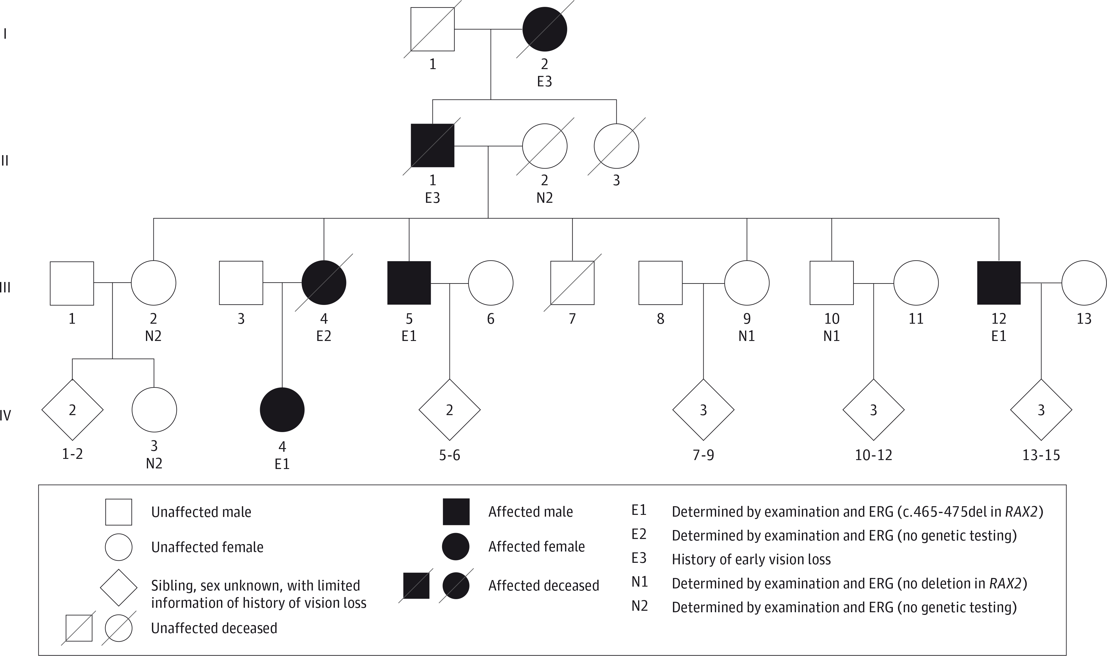
Importance: The patients evaluated in this study, to our knowledge, represent the first complete clinical description of a family with an autosomal dominant inheritance pattern of retinal dystrophy associated with a novel mutation in RAX2.
Objectives: To clinically evaluate 4 patients and 5 unaffected family members, characterize the disease phenotype over time, and identify the associated genetic mutation.
Design, setting, and participants: A prospective, longitudinal, observational, case-series analysis of 9 members of an affected family at the Casey Eye Institute, Oregon Health and Science University, Portland. The dates of the study were from July 31, 1992, to August 11, 2014.
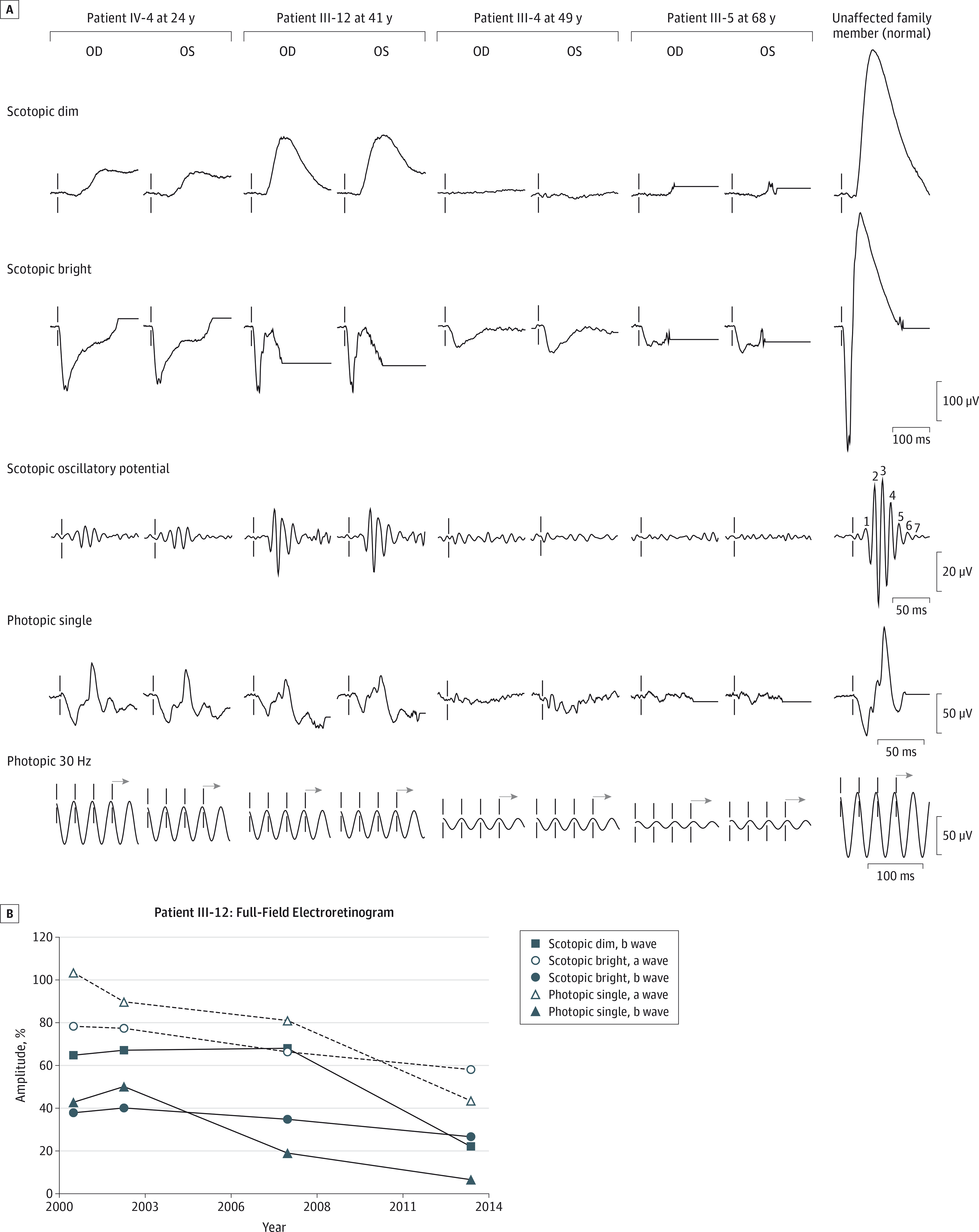
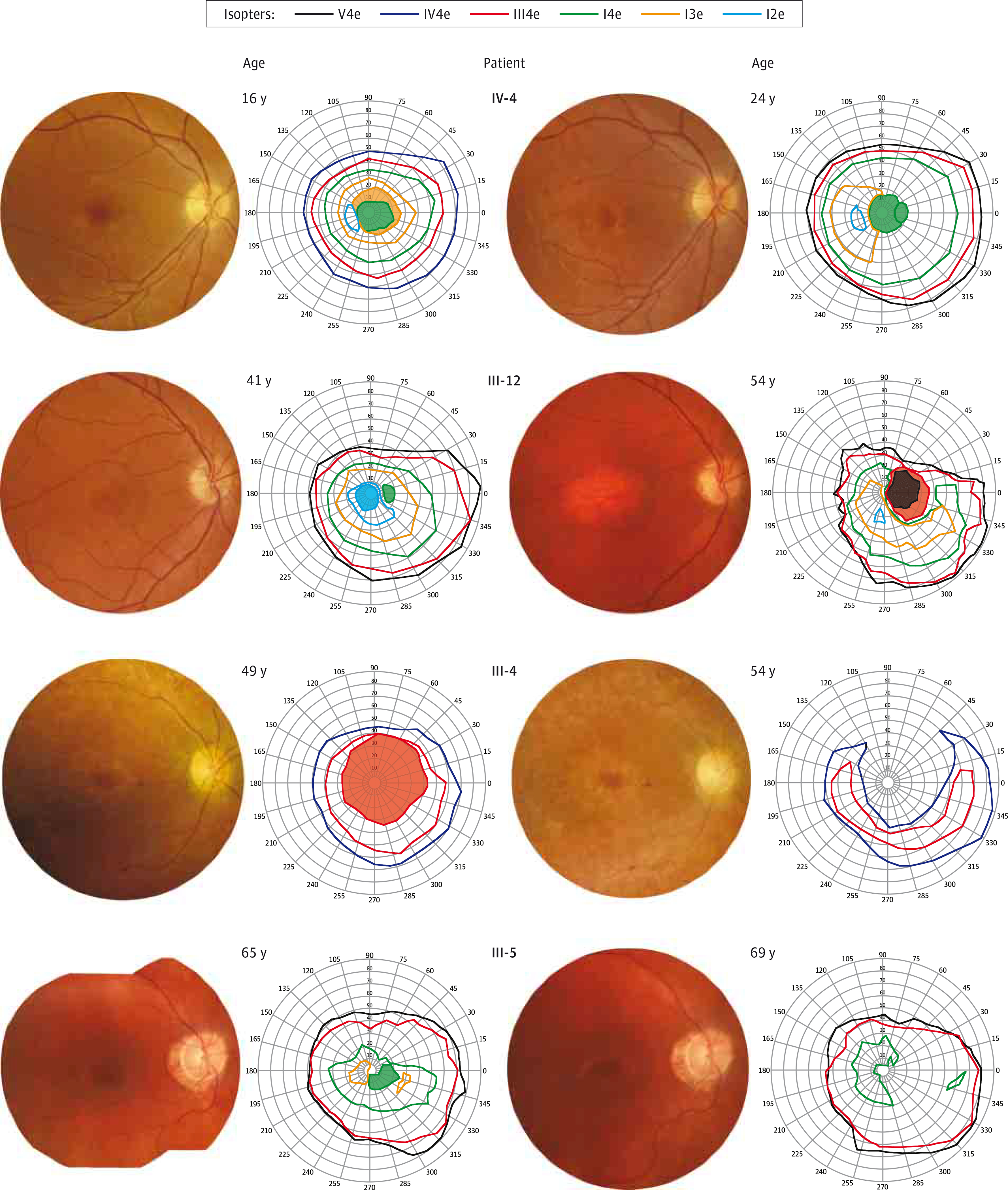
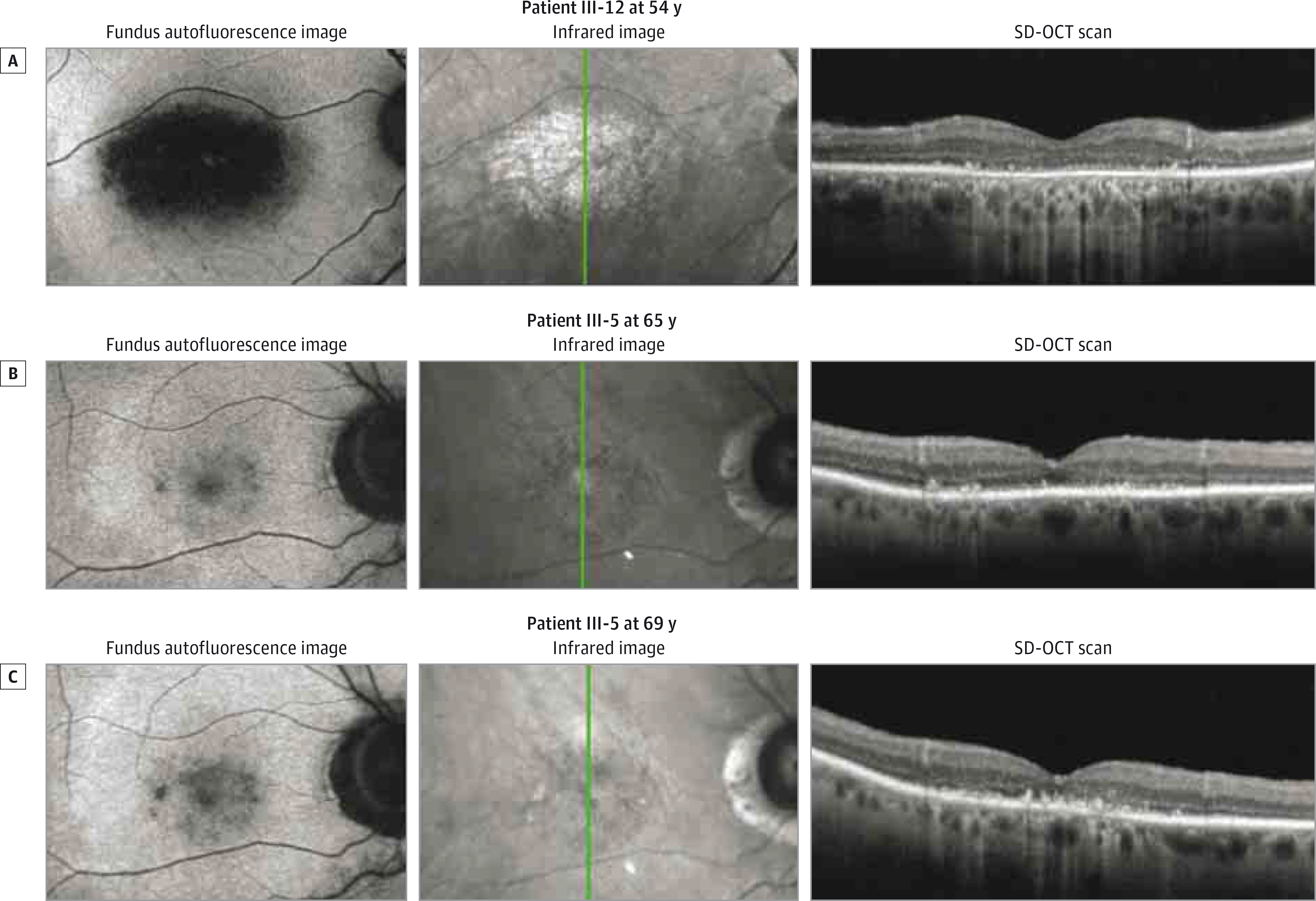
Interventions: Clinical evaluations included eye examination, color fundus photography, autofluorescence imaging, spectral-domain optical coherence tomography, kinetic visual field testing, and electroretinography. Genetic mutation screening was performed with next-generation sequencing, and identified mutations were confirmed with Sanger sequencing.
Main outcomes and measures: Clinical diagnosis and longitudinal characterization of retinal dystrophy and identification of genetic mutation.
Results: Six members of the family were identified as having retinal dystrophy (4 were examined, and 3 were genetically tested). Five unaffected family members were clinically evaluated (2 were genetically tested). The age at onset of retinal dystrophy was variable. All affected individuals presented with declining visual acuity, central scotomas, waxy disc pallor, attenuated vasculature, small yellow macular deposits and/or macular pigment mottling, and abnormal electroretinograms demonstrating mixed cone and rod dysfunction and a scotopic electronegative response to bright flashes. There were no other causes of an electronegative electroretinogram identified in any of the affected patients. Genetic testing revealed, to our knowledge, a novel frameshift heterozygous mutation in RAX2 in the patients with retinal dystrophy.
Conclusions and relevance: A frameshift heterozygous mutation in RAX2 inherited in an autosomal dominant fashion was associated with mixed cone and rod dysfunction. Among the patients, there was variability in the age at onset and in the specific pattern of photoreceptor dysfunction, but the clinical course was nevertheless slowly progressive. Screening for RAX2 mutation could provide prognostic value for patients and families with scotopic electronegative responses to bright flashes.
Figures
Similar articles
-
Electrophysiologic and phenotypic features of an autosomal cone-rod dystrophy caused by a novel CRX mutation.Ophthalmology. 2002 Oct;109(10):1862-70. doi: 10.1016/s0161-6420(02)01187-9. Ophthalmology. 2002. PMID: 12359607
-
Childhood cone-rod dystrophy with macular cystic degeneration from recessive CRB1 mutation.Ophthalmic Genet. 2014 Sep;35(3):130-7. doi: 10.3109/13816810.2013.804097. Epub 2013 Jun 14. Ophthalmic Genet. 2014. PMID: 23767994
-
Next-generation sequencing to solve complex inherited retinal dystrophy: A case series of multiple genes contributing to disease in extended families.Mol Vis. 2017 Jul 20;23:470-481. eCollection 2017. Mol Vis. 2017. PMID: 28761320 Free PMC article.
-
Genotype-phenotype correlation in a German family with a novel complex CRX mutation extending the open reading frame.Ophthalmology. 2007 Jul;114(7):1348-1357.e1. doi: 10.1016/j.ophtha.2006.10.034. Epub 2007 Feb 22. Ophthalmology. 2007. PMID: 17320181
-
Prevalence and novelty of PRPF31 mutations in French autosomal dominant rod-cone dystrophy patients and a review of published reports.BMC Med Genet. 2010 Oct 12;11:145. doi: 10.1186/1471-2350-11-145. BMC Med Genet. 2010. PMID: 20939871 Free PMC article. Review.
Cited by
-
The rax homeobox gene is mutated in the eyeless axolotl, Ambystoma mexicanum.Dev Dyn. 2021 Jun;250(6):807-821. doi: 10.1002/dvdy.246. Epub 2020 Sep 17. Dev Dyn. 2021. PMID: 32864847 Free PMC article.
-
PABPC1-induced stabilization of BDNF-AS inhibits malignant progression of glioblastoma cells through STAU1-mediated decay.Cell Death Dis. 2020 Feb 3;11(2):81. doi: 10.1038/s41419-020-2267-9. Cell Death Dis. 2020. PMID: 32015336 Free PMC article.
-
Biallelic sequence and structural variants in RAX2 are a novel cause for autosomal recessive inherited retinal disease.Genet Med. 2019 Jun;21(6):1319-1329. doi: 10.1038/s41436-018-0345-5. Epub 2018 Oct 31. Genet Med. 2019. PMID: 30377383 Free PMC article.
-
Origin and evolution of the Rax homeobox gene by comprehensive evolutionary analysis.FEBS Open Bio. 2020 Apr;10(4):657-673. doi: 10.1002/2211-5463.12832. Epub 2020 Mar 19. FEBS Open Bio. 2020. PMID: 32144893 Free PMC article.
-
Negative electroretinograms: genetic and acquired causes, diagnostic approaches and physiological insights.Eye (Lond). 2021 Sep;35(9):2419-2437. doi: 10.1038/s41433-021-01604-z. Epub 2021 Jun 14. Eye (Lond). 2021. PMID: 34127841 Free PMC article. Review.
References
-
- Michaelides M, Hardcastle AJ, Hunt DM, Moore AT. Progressive cone and cone-rod dystrophies: phenotypes and underlying molecular genetic basis. Surv Ophthalmol. 2006;51(3): 232–258. - PubMed
-
- Wang QL, Chen S, Esumi N, et al. QRX, a novel homeobox gene, modulates photoreceptor gene expression. Hum Mol Genet. 2004;13(10): 1025–1040. - PubMed
-
- Marmor MF, Fulton AB, Holder GE, Miyake Y, Brigell M, Bach M; International Society for Clinical Electrophysiology of Vision. ISCEV Standard for full-field clinical electroretinography (2008 update). Doc Ophthalmol. 2009;118(1):69–77. - PubMed
-
- Oh KT, Weleber RG, Stone EM, Oh DM, Rosenow J, Billingslea AM. Electroretinographic findings in patients with Stargardt disease and fundus flavimaculatus. Retina. 2004;24(6): 920–928. - PubMed
-
- van der Tweel LH, Estevez O. Analytical techniques. In: Heckenlively J, Arden GB, eds. Principles and Practice of Clinical Electrophysiology of Vision. 2nd ed. Cambridge, MA: MIT Press; 2006: 439–459.
Publication types
MeSH terms
Substances
Grants and funding
LinkOut - more resources
Full Text Sources
Molecular Biology Databases
