. 2015 Mar 18;85(6):1212-26.
doi: 10.1016/j.neuron.2015.02.026.

Mutant huntingtin downregulates myelin regulatory factor-mediated myelin gene expression and affects mature oligodendrocytes

Affiliations

Mutant huntingtin downregulates myelin regulatory factor-mediated myelin gene expression and affects mature oligodendrocytes

Brenda Huang et al. Neuron. .

Abstract

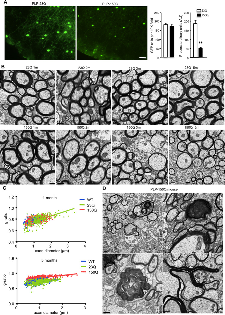
Growing evidence indicates that non-neuronal mutant huntingtin toxicity plays an important role in Huntington's disease (HD); however, whether and how mutant huntingtin affects oligodendrocytes, which are vitally important for neural function and axonal integrity, remains unclear. We first verified the presence of mutant huntingtin in oligodendrocytes in HD140Q knockin mice. We then established transgenic mice (PLP-150Q) that selectively express mutant huntingtin in oligodendrocytes. PLP-150Q mice show progressive neurological symptoms and early death, as well as age-dependent demyelination and reduced expression of myelin genes that are downstream of myelin regulatory factor (MYRF or MRF), a transcriptional regulator that specifically activates and maintains the expression of myelin genes in mature oligodendrocytes. Consistently, mutant huntingtin binds abnormally to MYRF and affects its transcription activity. Our findings suggest that dysfunction of mature oligodendrocytes is involved in HD pathogenesis and may also make a good therapeutic target.


Figures

Figure 1
Figure 1. Expression of mutant Htt in oligodendrocytes in HD 140Q knock-in mouse brain
(A) PLP-GFP mice were crossed with HD 140Q KI mice, and the brains of the crossed mice (PLP-GFP/KI) at one year of age were examined via immunofluorescent staining with anti-Htt (red). Arrows: GFP-positive oligodendrocytes (green) also express mutant Htt (red), though neuronal cells show more abundant Htt staining. (B) In the brain cortex sections that reveal processes of GFP-positive oligodendrocytes, GFP-positive processes are shorter in KI mouse brains. Length is reported in arbitrary units (AU). (C) Quantitative analysis of processes of GFP-positive oligodendrocytes in WT and KI mouse brain cortex. *** p<0.001. (3 mice per group). Scale bars: (A), 10 µm; (B), 5 µm.
Figure 2
Figure 2. Generation of PLP-HD transgenic mice
(A) DNA construct of the vector used to generate transgenic mice expressing N-terminal Htt (1–212 amino acids) with 23Q or 150Q under the control of the PLP promoter. (B) EM48 western blot analysis of transgenic mouse brain regions (brain stem and striatum) expressing transgenic mutant Htt (150Q4a line) and normal Htt (23Q-2 and 23Q-4). Aggregated Htt in the stacking gel is indicated. (C) 1C2 western blot analysis of multiple PLP-150Q lines showing mutant Htt expression in different brain tissues, but not in peripheral tissues. (D) mEM48 immunohistochemical staining of the striatum of PLP-150Q mice at 1, 2, and 3 months of age showing an increased accumulation of mutant Htt in the older mice. Right panels show high magnification micrographs. Scale bars: 10 µm.
Figure 3
Figure 3. Selective expression of mutant Htt in oligodendrocytes in adult PLP-150Q mice
(A) PLP-150Q mice were crossed with PLP-GFP mice, resulting in PLP-150Q/GFP mice in which GFP-positive oligodendrocytes (arrows) express mutant Htt. Immunostaining with mEM48 (red) showing that mutant Htt is enriched in the GFP-positive cells in the white matter (corpus callosum) and also localized in the nucleus. (B) Double immunocytochemical staining with antibodies to Htt (mEM48) and Olig2 showing the selective expression of mutant Htt in oligodendrocytes in in the striatum of 3-month-old PLP-150Q mouse. (C) Double staining with antibodies to GFAP and NeuN revealed that mutant Htt is expressed in GFP-positive oligodendrocytes, but not in GFAP-positive astrocytes or NeuN-positive neuronal cells. Scale bars: (A), 40 µm; (B, C): 10 µm.
Figure 4
Figure 4. Progressive neurological symptoms of PLP-150Q mice
(A) Representative photos of PLP-23Q and PLP-150Q mice, 6 months old. (B) Age-dependent loss of body weight in PLP-150Q mice; at least 15 animals per group. (C) Early death of PLP-150Q mice. The survival plot shows that PLP-150Q mice die at the age of 5–10 months, n=15. (D) Age-dependent worsening of rotarod performance of PLP-150Q mice compared with wild-type and PLP-23Q control mouse lines. At least 15 animals per genotype were examined. (E) Motor deficits in different PLP-150Q mouse lines compared with the PLP-23Q mouse line, n=5. (F) Locomotor activity was recorded for 24 hours. Statistical analysis shows reduced activity of PLP-150Q mice during the dark cycle, n=8–9, 2–3 months of age. (G) Increased susceptibility of PLP-150Q mice to flurothyl-induced seizures. At least 10 animals per genotype, 3–6 months of age. In (A, D-G), data are mean ± SEM. (E) and (G), one-way ANOVA, p<0.05. (B) and (F), two-way ANOVA, p<0.05. In (B), * significant compared to PLP-23Q; ** significant compared to PLP-23Q and WT.
Figure 5
Figure 5. Demyelination and axonal degeneration in PLP-150Q mice
(A) Loss of oligodendrocyte processes in the brain cortex in PLP-150Q mouse. Quantitative analysis of the number of GFP-positive oligodendrocytes and process length is shown in the right panel. Oligodendrocytes in PLP-150Q mouse brain have significantly shorter processes than PLP-23Q. Process length is reported in arbitrary units (AU). Data are mean ± SEM. ** P<0.01. (B) Electron microscopic graphs of the striatum of PLP-23Q and PLP-150Q mice at 1, 2, 3, and 5 months of age. When PLP-150Q mice become old (3 and 5 months), they show demyelination compared with one-month-old PLP-150Q mice and the age-matched PLP-23Q mice. Scale bars: 0.5 µm. (C) G-ratios were calculated and plotted against axon diameter with linear regression. G-ratio is significantly increased in PLP-150Q striatum (g=0.7568±0.0068) compared to age-matched WT and PLP-23Q (g=0.6315±0.0099 and g=0.6083±.0095, respectively). One-way ANOVA, p<0.05. At least 327 axons per genotype were examined. (D) Axonal degeneration and demyelination were also seen in PLP-150Q mice at the age of 5 months. Scale bar: 0.5 µm.
Figure 6
Figure 6. Reduced expression of myelin genes in PLP-150Q mice
(A) Western blotting showing up-regulation of multiple myelin proteins (MBP, CNP, MOBP and MOG) in the mouse brains during postnatal days, which is not affected by mutant Htt. (B) Western blotting showing the age-dependent decrease of myelin proteins (MBP, CNP, MOBP and MOG) in the brain stem of adult PLP-150Q, but not PLP-23Q and WT, mice. Transcription factor MYRF for the myelin genes and oligodendroglial lineage marker olig-2 remain unchanged in PLP-150Q mouse brain. (C) Densitometric analysis of the relative levels of myelin proteins (ratio of myelin protein to GAPDH) in (B). (D) qPCR of MBP transcripts verified that MBP mRNA levels are reduced in PLP-150Q mouse brain (n=3/genotype). (A–D): n=3 per genotype and age. Data are mean ± SEM. Two-way ANOVA, p<0.05. (E, F) qPCR (E) and Western blot (F) analysis of the expression of MBP in the brain cortex of HD patients and control individuals (n=3 each group). (G) The relative levels of MBP were quantified by measuring the ratios of MBP to GAPDH on the western blots in (F). * p< 0.05; ** p<0.01.
Figure 7
Figure 7. Interaction of mutant Htt with MYRF
(A) Comparison of in vitro synthesized and transfected MYRF showing that transfected full-length MYRF is cleaved to N-terminal MYRF (nMYRF) in cells. (B) Inhibition of MG132 markedly increased the levels of transfected full-length MYRF and nMYRF, suggesting that the ubiquitin-proteasome system degrades them. (C) Immunoprecipitation of mutant Htt from HD KI mouse brain selectively co-precipitates N-terminal MYRF (nMYRF). WT and IgG IP served as controls. (D) Immunoprecipitation of mutant Htt via mEM48 from PLP-150Q transgenic mouse brain also selectively co-precipitates N-terminal MYRF (nMYRF). Htt-150Q was preferentially labeled by 1C2 antibody on western blot (left panel). More nMYRF than full-length MYRF was precipitated with mutant Htt (right upper panel). Anti-GAPDH was also used to probe the immunoprecipitates (right low panel). (E) Expression of MYRF, nMYRF (1–635 aa) and cMYRF (616–1151 aa) in HEK293 cells. MYRF and nMYRF were detected with anti-myc, whereas cMYRF was detected by anti-flag. (F) Transfection of nMYRF and cMYRF into HEK293 cells revealed that only nMYRF is localized in the nucleus, supporting its role in gene transcription. Scale bars: 20 µm.
Figure 8
Figure 8. Mutant Htt binds N-terminal MYRF and inhibits its transcription activity
(A) Co-transfection of nMYRF (left panel) with N-terminal Htt containing 23Q or 150Q into HEK293 cells and immunoprecipitation of nMYRF. More Htt-150Q binds nMYRF than Htt-23Q. Co-transfection of cMYRF (right panel) with N-terminal Htt containing 23Q or 150Q into HEK293 cells showing no interaction between cMYRF and Htt. (B) The ratio of precipitated to input in (a) is shown. (C) GST-nMYRF and GST were expressed and purified to incubate with in vitro synthesized N-terminal Htt containing 23Q and 150Q also demonstrating that mutant Htt directly binds more MYRF. (D) MBP promoter was co-expressed with MYRF and Htt to assess its transcription activity via luciferase assay. MYRF markedly promotes MBP promoter activity. Co-expression of N-terminal Htt with 23Q did not significantly (p>0.05) affect the MBP promoter activity, whereas Htt-150Q significantly (p<0.001) reduces the MBP promoter activity. * p<0.05; *** p<0.001. (E) Expression of nMYRF, but not cMYRF, could rescue the inhibitory effect of mutant Htt on the MBP promoter activity. * p<0.05; ** p<0.01, *** p<0.001. (F) qPCR quantification of the MBP promoter DNAs associated with Myc-nMYRF that was immunoprecipitated by anti-Myc in ChIP assay. The results were obtained from three independent experiments. ** p=0.019. (G) A proposed model for the effect of mutant Htt in oligodendrocytes. According to recent studies (Bujalka et al., 2013; Li et al., 2013), full-length MYRF is self-cleaved to N-terminal MYRF (nMYRF), which is dissociated from ER and translocalized to the nucleus to activate the expression of myelin genes. In HD, the accumulation of N-terminal mutant Htt in the nucleus can lead to the abnormal binding of nMYRF and affects its transcription activity, leading to reduced expression of myelin genes and oligodendrocyte dysfunction.

References

    1. Bankston AN, Mandler MD, Feng Y. Oligodendroglia and neurotrophic factors in neurodegeneration. Neurosci Bull. 2013;29:216–228. - PMC - PubMed
    1. Bartzokis G, Lu PH, Tishler TA, Fong SM, Oluwadara B, Finn JP, Huang D, Bordelon Y, Mintz J, Perlman S. Myelin breakdown and iron changes in Huntington's disease: pathogenesis and treatment implications. Neurochem Res. 2007;32:1655–1664. - PubMed
    1. Bohanna I, Georgiou-Karistianis N, Sritharan A, Asadi H, Johnston L, Churchyard A, Egan G. Diffusion tensor imaging in Huntington's disease reveals distinct patterns of white matter degeneration associated with motor and cognitive deficits. Brain Imaging Behav. 2011;5:171–180. - PubMed
    1. Boulanger JJ, Messier C. From precursors to myelinating oligodendrocytes: Contribution of intrinsic and extrinsic factors to white matter plasticity in the adult brain. Neuroscience. 2014;269C:343–366. - PubMed
    1. Bradford J, Shin JY, Roberts M, Wang CE, Li XJ, Li S. Expression of mutant huntingtin in mouse brain astrocytes causes age-dependent neurological symptoms. Proc Natl Acad Sci U S A. 2009;106:22480–22485. - PMC - PubMed

Publication types