. 2015 Mar 19;6(3):e1700.
doi: 10.1038/cddis.2015.67.

Long noncoding RNA lincRNA-p21 is the major mediator of UVB-induced and p53-dependent apoptosis in keratinocytes

Affiliations

Long noncoding RNA lincRNA-p21 is the major mediator of UVB-induced and p53-dependent apoptosis in keratinocytes

J R Hall et al. Cell Death Dis. .

Abstract

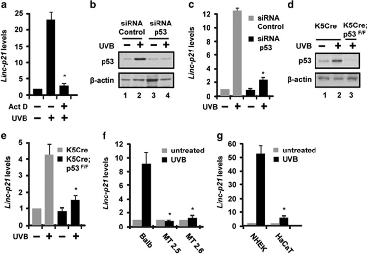
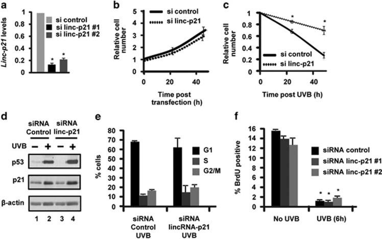
LincRNA-p21 is a long noncoding RNA and a transcriptional target of p53 and HIF-1α. LincRNA-p21 regulates gene expression in cis and trans, mRNA translation, protein stability, the Warburg effect, and p53-dependent apoptosis and cell cycle arrest in doxorubicin-treated mouse embryo fibroblasts. p53 plays a key role in the response of skin keratinocytes to UVB-induced DNA damage by inducing cell cycle arrest and apoptosis. In skin cancer development, UVB-induced mutation of p53 allows keratinocytes upon successive UVB exposures to evade apoptosis and cell cycle arrest. We hypothesized that lincRNA-p21 has a key functional role in UVB-induced apoptosis and/or cell cycle arrest in keratinocytes and loss of lincRNA-p21 function results in the evasion of apoptosis and/or cell cycle arrest. We observed that lincRNA-p21 transcripts are highly inducible by UVB in mouse and human keratinocytes in culture and in mouse skin in vivo. LincRNA-p21 is regulated at the transcriptional level in response to UVB, and the UVB induction of lincRNA-p21 in keratinocytes and in vivo in mouse epidermis is primarily through a p53-dependent pathway. Knockdown of lincRNA-p21 blocked UVB-induced apoptosis in mouse and human keratinocytes, and lincRNA-p21 was responsible for the majority of UVB-induced and p53-mediated apoptosis in keratinocytes. Knockdown of lincRNA-p21 had no effect on cell proliferation in untreated or UVB-treated keratinocytes. An early event in skin cancer is the mutation of a single p53 allele. We observed that a mutant p53(+/R172H) allele expressed in mouse epidermis (K5Cre(+/tg);LSLp53(+/R172H)) showed a significant dominant-negative inhibitory effect on UVB-induced lincRNA-p21 transcription and apoptosis in epidermis. We conclude lincRNA-p21 is highly inducible by UVB and has a key role in triggering UVB-induced apoptotic death. We propose that the mutation of a single p53 allele provides a pro-oncogenic function early in skin cancer development through a dominant inhibitory effect on UVB-induced lincRNA-p21 expression and the subsequent evasion of UVB-induced apoptosis.


Figures

Figure 1
Figure 1
LincRNA-p21 (Linc-p21) transcripts are highly inducible by UVB in mouse and human keratinocytes in culture and in mouse skin. (a) Balb/MK2 mouse keratinocytes were exposed to UVB, collected 8 h later and immunoblot analysis conducted. (b) Balb/MK2 mouse keratinocytes were exposed to the indicated doses of UVB, collected 16 h later and lincRNA-p21 transcript levels measured. (c) Balb/MK2 cells were exposed to 10 mJ/cm2 UVB and lincRNA-p21 transcript levels measured at the indicated times. (d) NHEK cells were exposed to the indicated doses of UVB, collected 16 h later and lincRNA-p21 transcripts measured. (e) NHEK cells were exposed to 10 mJ/cm2 UVB and lincRNA-p21 transcripts measured at the indicated times. (f) SKH-1 mice (three mice per time point) were exposed to 100 or 200 mJ/cm2 UVB, epidermis was collected at 9 h and lincRNA-p21 levels measured. (g) SKH-1 mice (three mice per group) were treated with 100 mJ/cm2 and lincRNA-p21 transcripts measured at the indicated times. LincRNA-p21 levels were measured by TaqMan real-time PCR (Ct value at peak post UVB=26–29 cycles using 25 ng template for all experiments). LincRNA-p21 was normalized to β-actin. Data are expressed as the mean ±S.D. N≥3, *P <0.05 significantly different compared with time 0 using student t-test
Figure 2
Figure 2
LincRNA-p21 is regulated at the transcriptional level and is p53-dependent in mouse and human keratinocytes and mouse skin in vivo. (a) Balb/MK2 keratinocytes were treated with actinomycin D, exposed to 10 mJ/cm2 UVB, collected 12 h later and lincRNA-p21 transcripts measured. (b) Balb/MK2 cells were transfected with siRNA to p53 or control siRNA, and 48 h post transfection, cells were exposed to 10 mJ/cm2 UVB, collected 12 h later and p53 protein levels were measured by immunoblot analysis. (c) Balb/MK2 cells were transfected with siRNA to p53 or control siRNA, and 48 h post transfection, cells were exposed to 10 mJ/cm2 UVB, collected 12 h later and lincRNA-p21 transcripts measured. (d) K5Cre+/tg and K5Cre+/tg;p53flox/flox SKH-1 mice were treated with 200 mJ/cm2 UVB and epidermis collected 9 h later and p53 protein levels were measured by immunoblot analysis. (e) K5Cre+/tg and K5Cre+/tg;p53flox/flox mice were treated with 200 mJ/cm2 UVB and epidermis collected 9 h later and lincRNA-p21 transcripts measured. (f) MT2.5, MT2.6 and Balb/MK2 cells were exposed to 10 mJ/cm2 UVB and collected 8 h later. (g) NHEK and HaCaT cells were exposed to 10 mJ/cm2 UVB and collected 12 h later. LincRNA-p21 transcript levels were determined by TaqMan real-time PCR. Expression of lincRNA-p21 was normalized to β-actin. Data expressed in a, c, e, f and g as the mean ±S.D. N ≥3, *P<0.05 significantly different compared to UVB exposed control as determined by the student t-test
Figure 3
Figure 3
LincRNA-p21 (linc-p21) does not regulate cell proliferation in UVB-treated keratinocytes. (a) Balb/MK2 cells were transfected with siRNA to lincRNA-p21 or control siRNA, and 48 h post transfection, cells were exposed to 10 mJ/cm2 UVB, collected 18 h later and lincRNA-p21 transcripts measured. (b) Control siRNA and lincRNA-p21 #1 knockdown Balb/MK2 cells were counted at 0, 24 and 48 h post transfection. (c) Control and lincRNA-p21 #1 knockdown Balb/MK2 cells were exposed to 10 mJ/cm2 UVB (48 h post transfection, t=0), and cells were counted 24 and 48 h after UVB (N=3 for each time point). (d) Control and lincRNA-p21 #1 knockdown Balb/MK2 cells were exposed to 10 mJ/cm2 UVB (48 h post transfection) and collected 18 h after UVB and immunoblot analysis conducted. (e) Control and lincRNA-p21 #1 knockdown Balb/MK2 cells were exposed to 10 mJ/cm2 UVB (48 h post transfection) and collected 24 h after UVB, PI-stained and analyzed by FACS. (f) Control and lincRNA-p21 #1 and #2 knockdown Balb/MK2 cells were exposed to 10 mJ/cm2 UVB (48 h post transfection) and pulse-labeled with BrdU for 1 h and collected 6 h after UVB. Cells were stained with BrdU-FITC and PI-stained and analyzed by FACS. Data expressed in a, b, c, e and f as the mean ±S.D. N ≥3, *P<0.05 significantly different compared with UVB-exposed control as determined by the student t-test
Figure 4
Figure 4
LincRNA-p21 (linc-p21) regulates apoptosis in UVB-treated keratinocytes. (a) Control and lincRNA-p21 #1 knockdown Balb/MK2 cells were exposed to 10 mJ/cm2 UVB (48 h post transfection) and collected at 0, 8 and 18 h after UVB exposure and immunoblot analysis for caspase 3 conducted; results shown are representative of duplicate experiments. (b) Control and lincRNA-p21 #1 knockdown Balb/MK2 cells were exposed to 10 mJ/cm2 UVB (48 h post transfection) and collected at 0, 6, 12, 18 and 24 h after UVB. Cells were stained with annexin-V and PI and analyzed by FACS. Similar results were obtained in three independent experiments. (c) Control and lincRNA-p21 #1 and #2 knockdown Balb/MK2 cells were exposed to 10 mJ/cm2 UVB (48 h post transfection) and 24 h after UVB. Cells were stained with annexin-V and PI and analyzed by FACS. Similar results were obtained in three independent experiments. (d) Control and lincRNA-p21 #1 and #2 knockdown Balb/MK2 cells were exposed to 10 mJ/cm2 and collected at 18 h post UVB and candidate gene expression examined by Taqman RT-PCR. (e) Control and lincRNA-p21 #1 and #2 knockdown Balb/MK2 cells were exposed to 10 mJ/cm2 and collected at 18 h post UVB and immunoblot analysis conducted. (f) NHEK cells were transfected with siRNA to human lincRNA-p21 or control siRNA, 48 h later exposed to 10 mJ/cm2 UVB, collected 18 h later and lincRNA-p21 levels measured. (g) Control and lincRNA-p21 knockdown NHEK cells were exposed to 10 mJ/cm2 UVB (48 h post transfection) and collected at 0 and 18 h after UVB. Cells were stained with annexin-V and PI and analyzed by FACS. Similar results were obtained in three independent experiments. (h) Control and lincRNA-p21 knockdown NHEK cells were exposed to 10 mJ/cm2 and collected at 18 h post UVB, and candidate gene expression examined. Data are expressed in c, d, f, g and h as the mean ±S.D. N ≥3, *P<0.05 significantly different compared with siRNA control as determined by the student t-test
Figure 5
Figure 5
Mutation of single p53 allele has a dominant-negative effect in vivo in mouse epidermis. (a) K5Cre+/tg, K5Cre+/tg;LSLp53+/R172H, K5Cre+/tg;p53+/flox and K5Cre+/tg;p53flox/flox SKH-1 mice were treated with 200 mJ/cm2 UVB and epidermal lincRNA-p21 transcripts measured 9 h post UVB treatment. (b) K5Cre+/tg, K5Cre+/tg;LSLp53+/R172H, K5Cre+/tg;p53+/flox and K5Cre+/tg;p53flox/flox SKH-1 mice were treated with 100 mJ/cm2 UVB and the number of apoptotic interfollicular basal epidermal keratinocytes/cm skin were scored at 9 h post UVB treatment. Data are expressed as the mean ±S.D. N≥3, *P <0.05 significantly different compared with UVB-treated K5Cre mice as determined by the student t-test

References

    1. Lander ES, Linton LM, Birren B, Nusbaum C, Zody MC, Baldwin J, et al. Initial sequencing and analysis of the human genome. Nature. 2001;409:860–921. - PubMed
    1. Bertone P, Stolc V, Royce TE, Rozowsky JS, Urban AE, Zhu X, et al. Global identification of human transcribed sequences with genome tiling arrays. Science. 2004;306:2242–2246. - PubMed
    1. Kapranov P, Cheng J, Dike S, Nix DA, Duttagupta R, Willingham AT, et al. RNA maps reveal new RNA classes and a possible function for pervasive transcription. Science. 2007;316:1484–1488. - PubMed
    1. Mattick JS. The genetic signatures of noncoding RNAs. PLoS Genet. 2009;5:e1000459. - PMC - PubMed
    1. Mercer TR, Gerhardt DJ, Dinger ME, Crawford J, Trapnell C, Jeddeloh JA, et al. Targeted RNA sequencing reveals the deep complexity of the human transcriptome. Nat Biotechnol. 2012;30:99–104. - PMC - PubMed

Publication types

MeSH terms

Substances