Clinical Trial
. 2015 Mar 19;10(3):e0113483.
doi: 10.1371/journal.pone.0113483. eCollection 2015.

A pilot study of omalizumab in eosinophilic esophagitis

Affiliations
Clinical Trial

A pilot study of omalizumab in eosinophilic esophagitis

Denise Loizou et al. PLoS One. .

Abstract

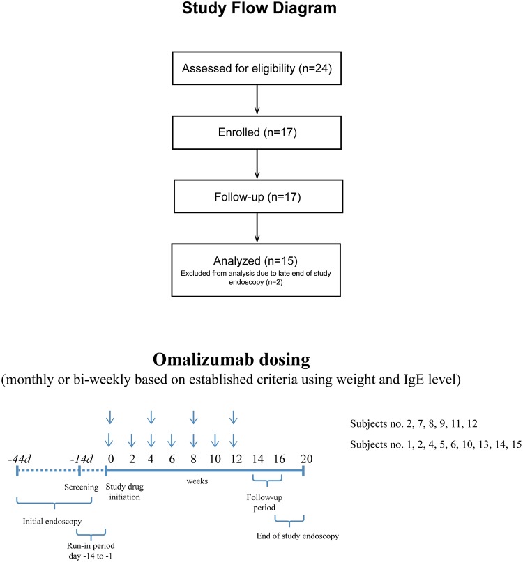
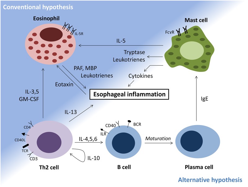
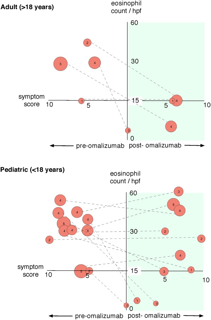
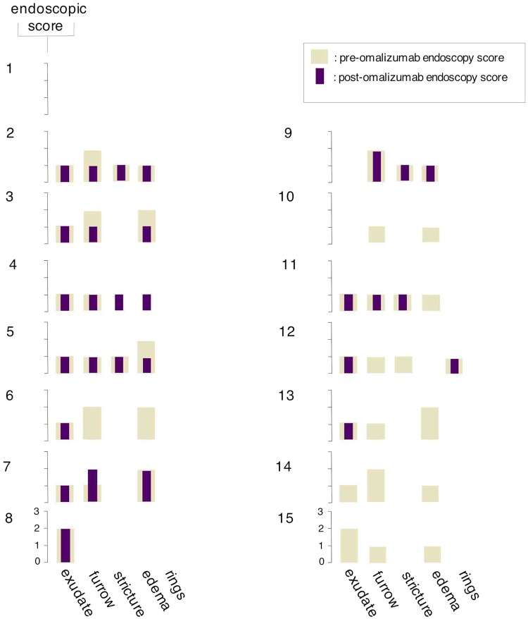
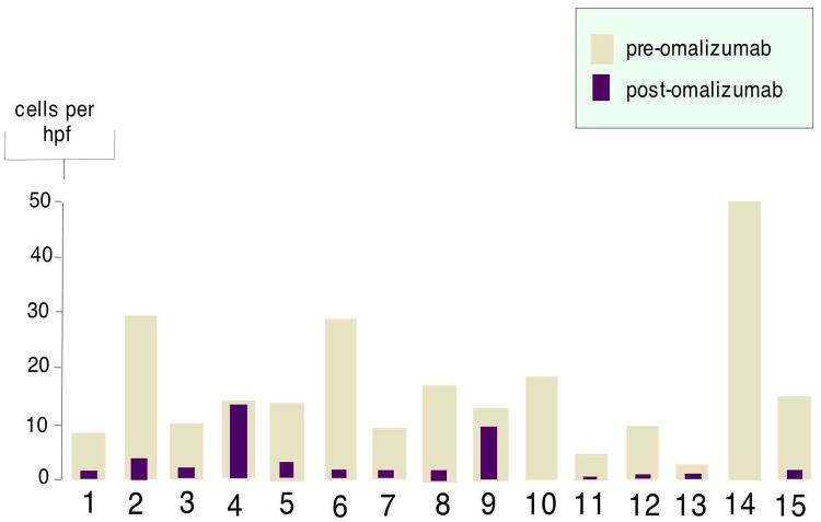
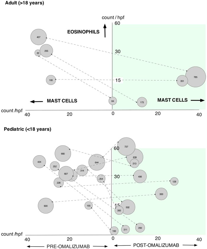
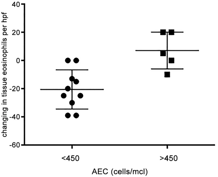
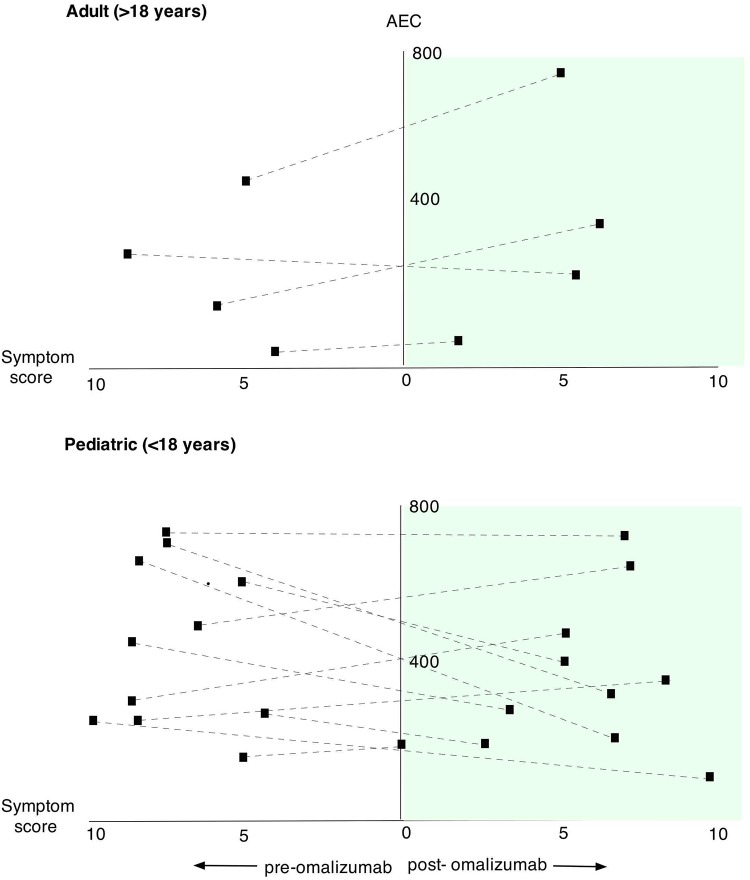
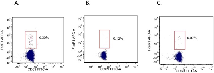
Eosinophilic disorders of the gastrointestinal tract are an emerging subset of immune pathologies within the spectrum of allergic inflammation. Eosinophilic Esophagitis (EoE), once considered a rare disease, is increasing in incidence, with a rate of over 1 in 10,000 in the US, for unknown reasons. The clinical management of EoE is challenging, thus there is an urgent need for understanding the etiology and pathophysiology of this eosinophilic disease to develop better therapeutic approaches. In this open label, single arm, unblinded study, we evaluated the effects of an anti-IgE treatment, omalizumab, on local inflammation in the esophagus and clinical correlates in patients with EoE. Omalizumab was administered for 12 weeks to 15 subjects with long standing EoE. There were no serious side effects from the treatment. Esophageal tissue inflammation was assessed both before and after therapy. After 3 months on omalizumab, although tissue Immunoglobulin E (IgE) levels were significantly reduced in all but two of the subjects, we found that full remission of EoE, which is defined as histologic and clinical improvement only in 33% of the patients. The decrease in tryptase-positive cells and eosinophils correlated significantly with the clinical outcome as measured by improvement in endoscopy and symptom scores, respectively. Omalizumab-induced remission of EoE was limited to subjects with low peripheral blood absolute eosinophil counts. These findings demonstrate that in a subset of EoE patients, IgE plays a role in the pathophysiology of the disease and that anti-IgE therapy with omalizumab may result in disease remission. Since this study is open label there is the potential for bias, hence the need for a larger double blind placebo controlled study. The data presented in this pilot study provides a foundation for proper patient selection to maximize clinical efficacy.

Trial registration: ClinicalTrials.gov NCT01040598.


Conflict of interest statement

Competing Interests: The study was funded by Genentech, Inc. and by O&O Alpan, LLC. Co-authors DL, EK-P, PH, JK-C, LN, SK, RL, MB, RG, NB, OG-A, AK & OA are employed by O&O ALPAN, LLC. Co-authors DL, RL, RG and OA are employed by Amerimmune. There are no other patents, products in development or marketed products to declare. This does not alter the authors' adherence to all the PLOS ONE policies on sharing data and materials.

Figures

Fig 1
Fig 1. Study flow diagram and design.
24 subjects consented to participate in the study, 17 enrolled and completed all study visits. A total of 15 subjects who fully met inclusion and exclusion criteria, completed all study visits and drug administration, and underwent pre-and post-treatment endoscopic examinations were included in the data analysis. After a 2-week screening period, subjects received omalizumab subcutaneously every 2 weeks (6 patients) or 4 weeks (9 patients) for a total of 12 weeks. Safety and efficacy continued to be monitored during the follow-up period (weeks 12 through 16). The end of study endoscopy was performed between week 20 and 24.
Fig 2
Fig 2. Mechanisms of eosinophilic esophagitis.
Conventional hypothesis: esophageal epithelial cells mediate eosinophil influx into esophagus. Alternative hypothesis: IgE mediated secretion of eosinophilic factors from mast cells.
Fig 3
Fig 3. Effect of omalizumab on esophageal eosinophil counts, symptom scores and overall endoscopic score.
The x-axis represents symptoms scores, the y-axis eosinophil counts, and right side of the graph represents post therapy with omalizumab. Individual patient data are shown as circles connected with a dashed line to allow comparison between pretreatment and post treatment. The size of the circle represents endoscopy score (also shown as a number inside the circle).
Fig 4
Fig 4. Endoscopic scoring of individual patients before and after therapy with omalizumab.
The y-axis represents symptoms scores; the x-axis represents endoscopic findings. The pale bar represent endoscopic score before therapy with omalizumab, and the purple bar represents endoscopic score 3 months after therapy with omalizumab. Numbers to the upper left of each graph shows individual patients. Endoscopic scores are graded as: 0–2 for exudates, 0–2 for furrows, 0–1 for stricture, 0–2 for edema and 0–3 for rings.
Fig 5
Fig 5. Effect of omalizumab on tissue IgE.
Immunohistochemical staining for IgE was performed at baseline and 3 months into omalizumab therapy. Positive stain is shown in per high power field.
Fig 6
Fig 6. Relationship between mast cells and eosinophils in the tissue and their comparison to blood eosinophil counts.
The x-axis represents mast cell count per high power field; the y-axis represents eosinophil counts per high power field. Left side of the graph is data before omalizumab therapy and right side of the graph after therapy. Individual patient data are shown as circles connected with a dashed line. The size of the circle represents peripheral blood absolute eosinophil count (also shown as a number inside the circle).
Fig 7
Fig 7. Change in tissue eosinophil counts/hpf in response to omalizumab therapy in patients with low and high peripheral blood absolute eosinophil counts.
Patients were classified as having high (>450 cell/mcl) and low (<450 cells/mcl) peripheral blood eosinophil counts and the tissue eosinophil counts/hpf before and after omalizumab were compared using t-test (p = 0.0027).
Fig 8
Fig 8. Comparison of blood AEC to symptom score.
The x-axis represents symptoms scores before (left) and after (right) omalizumab, the y-axis represents AEC in peripheral blood.
Fig 9
Fig 9. Eosinophil FcR1 expression in EoE.
Eosinophils were identified by a combination of FSC/SSA and expression of CD9 and CD16 as CD9+CD16- cells. Evaluation of FcεR1 and CD69 were compared versus IgG1 as an isotype control.

References

    1. Straumann A (2011) The natural history and complications of eosinophilic esophagitis. Thorac Surg Clin 21: 575–587. 10.1016/j.thorsurg.2011.09.004 - DOI - PubMed
    1. Straumann A, Schoepfer AM (2012) Therapeutic concepts in adult and paediatric eosinophilic oesophagitis. Nat Rev Gastroenterol Hepatol 9: 697–704. 10.1038/nrgastro.2012.182 - DOI - PubMed
    1. Spergel JM, Brown-Whitehorn TF, Cianferoni A, Shuker M, Wang ML, et al. (2012) Identification of causative foods in children with eosinophilic esophagitis treated with an elimination diet. J Allergy Clin Immunol 130: 461–467 e465. 10.1016/j.jaci.2012.05.021 - DOI - PubMed
    1. Wechsler ME, Fulkerson PC, Bochner BS, Gauvreau GM, Gleich GJ, et al. (2012) Novel targeted therapies for eosinophilic disorders. J Allergy Clin Immunol 130: 563–571. 10.1016/j.jaci.2012.07.027 - DOI - PMC - PubMed
    1. Mulder DJ, Justinich CJ (2011) Understanding eosinophilic esophagitis: the cellular and molecular mechanisms of an emerging disease. Mucosal Immunol 4: 139–147. 10.1038/mi.2010.88 - DOI - PubMed

Publication types

Associated data