. 2015 Mar 19;10(3):e0121070.
doi: 10.1371/journal.pone.0121070. eCollection 2015.

IL-4Rα-dependent alternative activation of macrophages is not decisive for Mycobacterium tuberculosis pathology and bacterial burden in mice

Affiliations

IL-4Rα-dependent alternative activation of macrophages is not decisive for Mycobacterium tuberculosis pathology and bacterial burden in mice

Reto Guler et al. PLoS One. .

Abstract

Classical activation of macrophages (caMph or M1) is crucial for host protection against Mycobacterium tuberculosis (Mtb) infection. Evidence suggests that IL-4/IL-13 alternatively activated macrophages (aaMph or M2) are exploited by Mtb to divert microbicidal functions of caMph. To define the functions of M2 macrophages during tuberculosis (TB), we infected mice deficient for IL-4 receptor α on macrophages (LysMcreIL-4Rα-/lox) with Mtb. We show that absence of IL-4Rα on macrophages does not play a major role during infection with Mtb H37Rv, or the clinical Beijing strain HN878. This was demonstrated by similar mortality, bacterial burden, histopathology and T cell proliferation between infected wild-type (WT) and LysMcreIL-4Rα-/lox mice. Interestingly, we observed no differences in the lung expression of inducible nitric oxide synthase (iNOS) and Arginase 1 (Arg1), well-established markers for M1/M2 macrophages among the Mtb-infected groups. Kinetic expression studies of IL-4/IL-13 activated bone marrow-derived macrophages (BMDM) infected with HN878, followed by gene set enrichment analysis, revealed that the MyD88 and IL-6, IL-10, G-CSF pathways are significantly enriched, but not the IL-4Rα driven pathway. Together, these results suggest that IL-4Rα-macrophages do not play a central role in TB disease progression.


Conflict of interest statement

Competing Interests: The authors have declared that no competing interests exist.

Figures

Fig 1
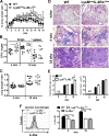
Fig 1. Increased acute bacterial burden and chronic pulmonary pathology in absence of IL-4Rα responsive macrophages following low-dose Mtb H37Rv infection
. Wild-type (BALB/c), IL-4Rα-/- and IL-4Rα macrophage cell-specific deficient mice (LysMcreIL-4Rα-/lox) were infected with Mtb H37Rv (100 CFU/mouse) by aerosol (n = 12–13 mice/group). (A) Percentages in body weight change are shown. (B) Mice were sacrificed at 4 and 18 weeks PI to determine bacterial loads in the lungs and spleen. (C) Lung weight indexes are shown. (D) At 0, 4 and 18 weeks PI, formalin-fixed lung sections were stained with H&E. Original magnification: 40X. (E) Lung sections of 5 mice per group were evaluated for pulmonary histopathology scores and quantification of total lesion sizes. N.D. = not detectable. (F) IL-4Rα expression was measured by flow cytometry on alveolar macrophages (SiglecF+CD11c+) at 0 and 18 weeks PI (*P < 0.05, **P < 0.01). Data shown are representative (A, D, E and F) and pooled (B, C) from two independent experiments.
Fig 2
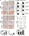
Fig 2. No major differences in expression of iNOS, Arg1, lung immune cell populations and T cell proliferation between wild-type and macrophage cell-specific IL-4Rα deficient mice following low-dose Mtb H37Rv infection (100 CFU/mouse).
(A) iNOS and Arg1 staining (brown colour) from lung sections collected at indicated times PI, original magnification: 40X. Lung sections from 5 mice/group were quantified. N.D. = not detectable. (B) iNOS and Arg1 expression on various immune cells were analysed by flow cytometry at 18 weeks PI (6–7 mice/group, *P < 0.05). (C) T cell proliferation with co-cultured CD11c-sorted macrophages from the lung tissue of naïve non-infected and mice infected with H37Rv (100 CFU/mouse) by aerosol at 4 and 18 weeks. Data shown in A is representative of two independent experiments and results obtained in B and C are from one experiment.
Fig 3
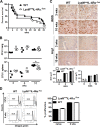
Fig 3. Similar mortality, inflammation, bacterial burden, Arg1/iNOS expression and T cell proliferation in wild-type and LysMcreIL-4Rα-/lox mice following high dose infection with Mtb H37Rv
. Wild-type (BALB/c) and IL-4Rα macrophage cell-specific deficient mice (LysMcreIL-4Rα-/lox) were infected intranasally with high dose of 104 CFU/mouse of Mtb H37Rv (n = 20/group). (A) Survival of infected mice was recorded weekly. (B) Individual bacterial titers (CFU/organ) with group medians are shown (*P < 0.05). (C) iNOS and Arg1 staining (brown colour) from lung sections collected at 3 and 10 weeks PI. Lung sections from 5 mice/group were quantified. Original magnification: 40X. N.D. = not detectable. (D) T cell proliferation with co-cultured CD11c-sorted macrophages from the lung tissue of naïve non-infected and 3 weeks infected mice. All data shown is representative of two independent experiments except for B which is representative of three independent experiments.
Fig 4
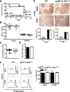
Fig 4. Macrophage cell-specific IL-4Rα deficient and wild-type mice displayed similar mortality, inflammation and T cell proliferation to hypervirulent Mtb HN878 infection
. Wild-type (BALB/c) and IL-4Rα macrophage cell-specific deficient mice (LysMcreIL-4Rα-/lox) were infected intranasally with 500 CFU/mouse of hypervirulent Mtb HN878. (A) Survival of infected mice was recorded weekly (n = 6–8/group). (B) Individual bacterial titers (CFU/organ), (C) lung weight indexes and (D) histopathology score with group medians are shown at 3 weeks PI (n = 11/group). (E) iNOS and Arg1 staining (brown colour) from lung sections collected at 3 weeks PI. Lung sections from 5 mice/group were quantified. Original magnification: 40X. N.D. = not detectable. (F) T cell proliferation with co-cultured CD11c-sorted macrophages from the lung tissue of naïve non-infected and mice infected with HN878 at 3 weeks PI. Data shown in A-E is representative of two independent experiments and results obtained in F is from one experiment.
Fig 5
Fig 5. MyD88 and IL-6, IL-10, G-CSF-dependent pathway genes are significantly enriched in HN878 infected vs. non-infected macrophages
. BMDM were stimulated with IL-4/IL-13 or left untreated. After 24 hours of stimulation, cells were infected with HN878. Total RNA was extracted at 4, 12 and 48 hours PI for microarray and GSEA analysis. Enrichment plots and heat maps for (A) MyD88, (B) IL-6, IL-10, G-CSF and (C) IL-4Rα pathway are shown. Enrichment analysis compared log2-fold changes in Mtb-infected samples vs. non-infected samples. The rows in heat maps are listed according to pre-ranking metric scores. Replicates shown are from two independent experiments.

References

    1. Benoit M, Desnues B, Mege JL. Macrophage polarization in bacterial infections. J Immunol. 2008. September 15;181(6):3733–9. - PubMed
    1. Martinez FO, Helming L, Gordon S. Alternative activation of macrophages: an immunologic functional perspective. Annu Rev Immunol. 2009;27:451–83. 10.1146/annurev.immunol.021908.132532 - DOI - PubMed
    1. Gordon S. Alternative activation of macrophages. Nat Rev Immunol. 2003. January;3(1):23–35. - PubMed
    1. Pathak SK, Basu S, Basu KK, Banerjee A, Pathak S, Bhattacharyya A, et al. Direct extracellular interaction between the early secreted antigen ESAT-6 of Mycobacterium tuberculosis and TLR2 inhibits TLR signaling in macrophages. Nat Immunol. 2007. June;8(6):610–8. - PubMed
    1. Ashenafi S, Aderaye G, Bekele A, Zewdie M, Aseffa G, Hoang AT, et al. Progression of clinical tuberculosis is associated with a Th2 immune response signature in combination with elevated levels of SOCS3. Clin Immunol. 2014. April;151(2):84–99. 10.1016/j.clim.2014.01.010 - DOI - PubMed

Publication types

MeSH terms

Substances

Associated data