. 2015 Mar 20:5:9261.
doi: 10.1038/srep09261.

Oligomerized, filamentous surface presentation of RANTES/CCL5 on vascular endothelial cells

Affiliations

Oligomerized, filamentous surface presentation of RANTES/CCL5 on vascular endothelial cells

Inger Øynebråten et al. Sci Rep. .

Abstract

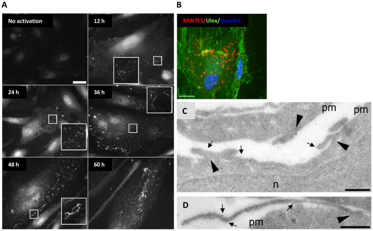
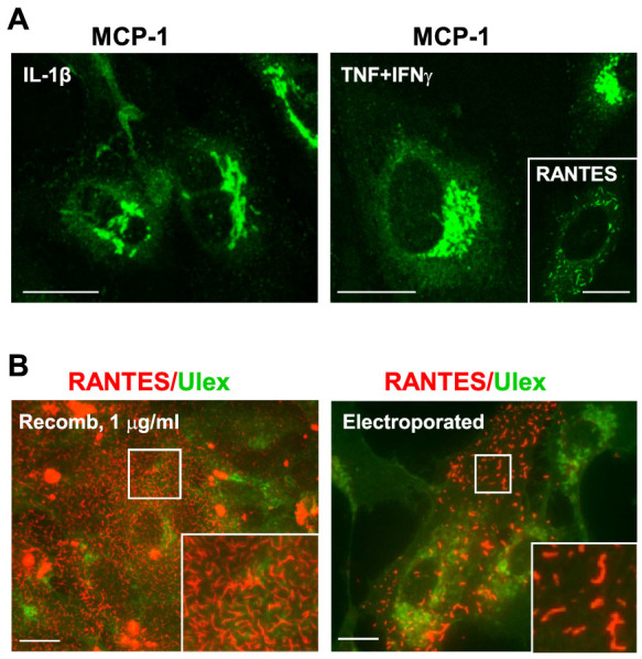
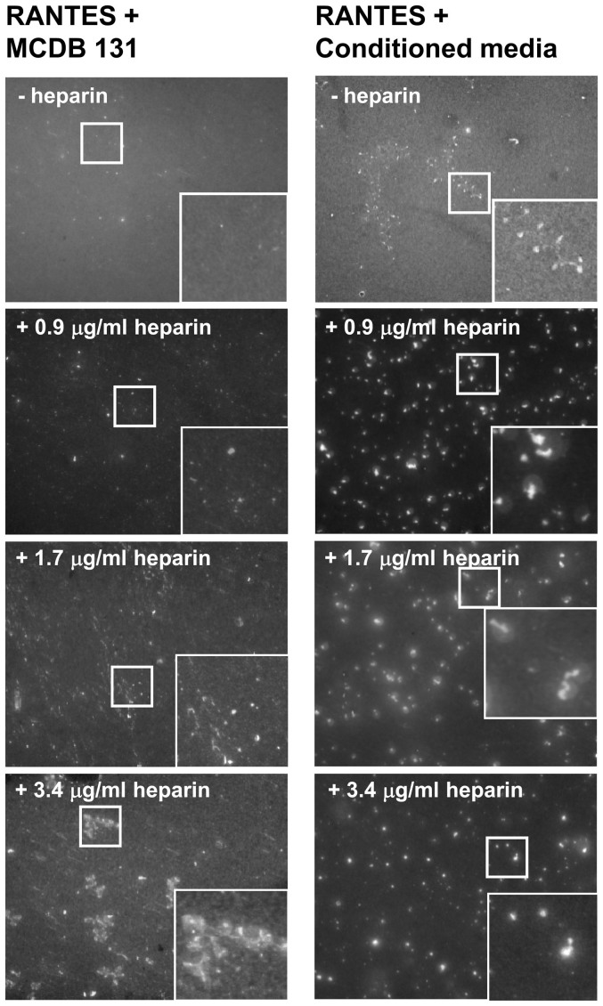
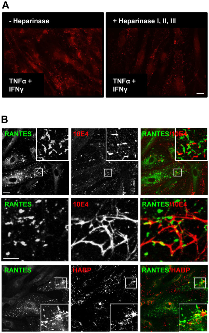
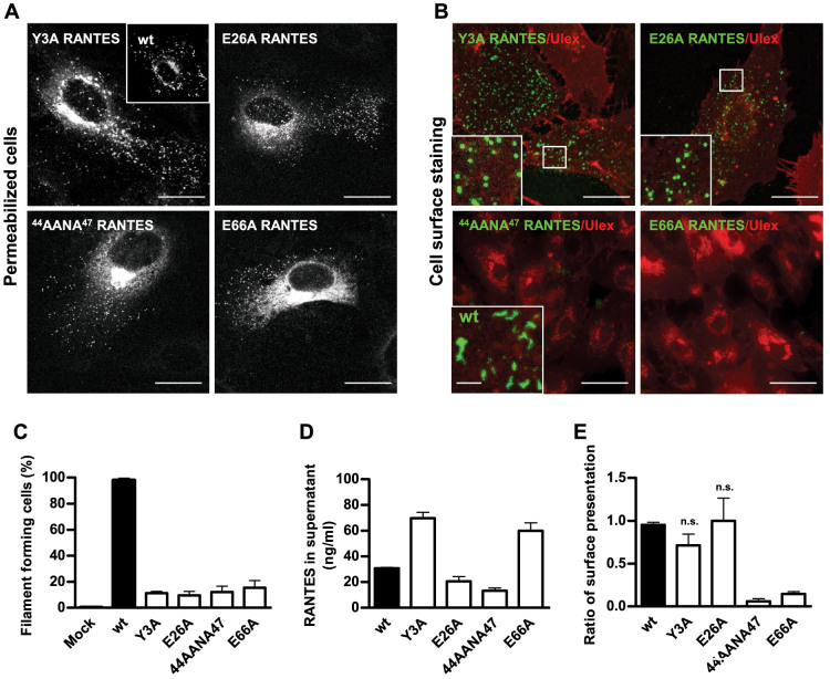
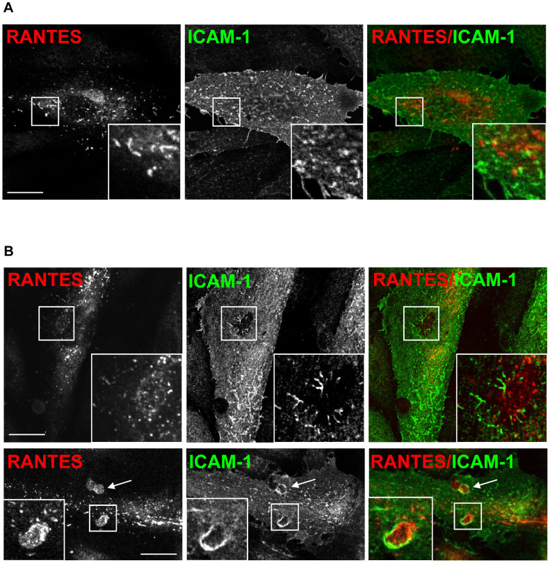
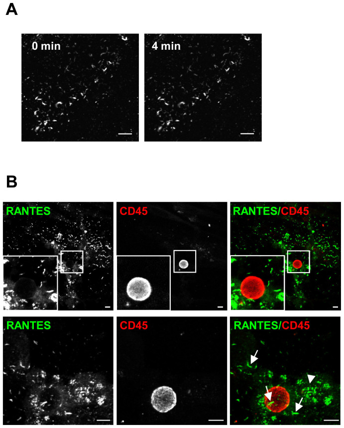
Vascular endothelial cells present luminal chemokines that arrest rolling leukocytes by activating integrins. It appears that several chemokines must form higher-order oligomers to elicit proper in vivo effects, as mutants restricted to forming dimers have lost the ability to recruit leukocytes to sites of inflammation. Here, we show for the first time that the chemokine RANTES/CCL5 binds to the surface of human endothelial cells in a regular filamentous pattern. Furthermore, the filaments bound to the surface in a heparan sulfate-dependent manner. By electron microscopy we observed labeling for RANTES on membrane projections as well as on the remaining plasma membrane. Mutant constructs of RANTES restricted either in binding to heparin, or in forming dimers or tetramers, appeared either in a granular, non-filamentous pattern or were not detectable on the cell surface. The RANTES filaments were also present after exposure to flow, suggesting that they can be present in vivo. Taken together with the lacking in vivo or in vitro effects of RANTES mutants, we suggest that the filamentous structures of RANTES may be of physiological importance in leukocyte recruitment.


Figures

Figure 1
Figure 1. RANTES organizes in filaments on the cell surface and the filament length increases with incubation time in the presence of TNFα and IFNγ.
(A) HUVECs were cultured in growth medium with 10 ng/ml TNFα and 1 ng/ml IFNγ for different time points (indicated in each image) before fixation and immunostaining with clone ID2/A12. Inserts show structure details at 3 × magnification. Scale bar, 10 μm. The images were acquired by widefield microscopy. (B) HUVECs were stimulated with 10 ng/ml TNFα and 1 ng/ml IFNγ for 30 h and then immunostained with a rabbit anti-RANTES antibody on ice to label only extracellular, surface associated RANTES. Ulex, a lectin, was utilized to label the surface of all HUVECs. Scale bar, 10 μm. Images were acquired by sequential scanning confocal microscopy. (C, D) HUVECs were stimulated with 10 ng/ml TNFα and 1 ng/ml IFNγ for 36 h and frozen for cryosectioning before immunogold detection of RANTES with a goat anti-RANTES antibody. The images show sections of the outer part of HUVECs, with membrane projections originating from the cell surface (arrowheads). Arrows indicate immunogold labelled RANTES. pm, plasma membrane; n, nucleus. Scale bars, 500 nm.
Figure 2
Figure 2. Filaments of RANTES form independently of TNFα and IFNγ-stimulation.
(A) HUVECs were stimulated with 1 ng/ml IL-1β, or 10 ng/ml TNFα in combination with 1 ng/ml IFNγ for 24 h and immunostained with an antibody towards MCP-1. The insert shows immunostaining of RANTES from the same experiment, in HUVECs stimulated with 10 ng/ml TNFα + 1 ng/ml IFNγ. (B) Left image; HUVECs were incubated with 1 μg/ml recombinant RANTES before fixation and immunostaining. Right image; HUVECs were not stimulated but electroporated with a DNA plasmid encoding RANTES before fixation and immunostaining of RANTES with a rabbit anti-RANTES antibody. Labelling with ulex (green) was included to visualize individual cells. Scale bars, 10 μm. All images were acquired by confocal microscopy.
Figure 3
Figure 3. RANTES is dependent on other molecules to form organized structures.
RANTES (1 μg/ml) was incubated either in cell growth medium without serum or in conditioned cell growth medium containing serum. The conditioned medium was harvested from unstimulated cultures of HUVECs. Heparin was added and its final concentration is indicated in each image. After 35 h, the samples were fixed and immunostained with a rabbit antibody toward RANTES. The images were acquired by widefield microscopy. Inserts show high magnification of squared areas. Scale bars, 10 μm.
Figure 4
Figure 4. RANTES is immobilized to the cell surface via heparan sulfate.
(A) HUVECs were stimulated for 48 h with 10 ng/ml TNFα + 1 ng/ml IFNγ before half of the samples were incubated with a mixture of heparinase I, II, and III (0.5 U/ml) for 2 h. Next, the cells were fixed and immunolabelled with clone ID2/A12 and analyzed by widefield microscopy. Scale bars, 50 μm. (B) HUVECs stimulated with 10 ng/ml TNFα and 1 ng/ml IFNγ were immunostained with antibodies towards RANTES (rabbit anti-RANTES antibody) and the heparan sulfate epitope 10E4. Alternatively, biotinylated hyaluronan binding protein (HABP) was used to label hyaluronan. The antibody towards 10E4 labelled elongated structures in HUVECs (upper panel) and long structures at cell borders or between cells (middle panel). Middle panel is a high magnification from an original 100 × picture. Biotinylated HABP labelled irregular clusters in HUVECs, and large, round structures between cells (lower panel). The samples were analyzed following sequential scanning confocal microscopy. Corner insets show high magnification of framed areas. Scale bars, 10 μm.
Figure 5
Figure 5. Oligomerization-deficient mutants of RANTES show distinct morphology and localization compared to the wild type (wt).
(A) HUVECs were electroporated with DNA plasmids encoding wtRANTES or oligomerization-deficient mutants and cultivated for 24 h before fixation. The cells were permeabilized to label RANTES present intracellularly and on the surface with rabbit anti-RANTES antibody. Images were acquired by confocal microscopy. Corner inset shows a cell electroporated with wtRANTES-encoding plasmid. Scale bars, 10 μm. (B) The experiment was performed as indicated in A, but labelling was performed on live HUVECs kept on ice to detect cell surface-associated RANTES with rabbit anti-RANTES antibody. Labelling with the lectin ulex was used to visualize individual cells. Images were acquired by sequential scanning confocal microscopy. Corner insets show high magnification of framed areas or high magnification of a cell electroporated with DNA encoding wtRANTES. Scale bars, 10 μm (wt, scale bar = 5 μm). (C) HUVECs were treated as described in A, and the number of RANTES-positive and filament-forming cells were counted. The graph presents mean values of percentage of filament-forming cells related to the total number of RANTES-positive cells. 55–80 cells were evaluated for each construct in one experiment (n = 3 experiments). Error bars indicate SEM. The mutants generated a significantly lower percentage of filament-forming cells than the wt, p < 0.0001. (D) HUVECs electroporated with DNA encoding the indicated constructs were incubated for 30 h before supernatants were harvested. The amount of RANTES in supernatants was quantified by ELISA utilizing recombinant RANTES as standard. The mutants were present in amounts that differed significantly from the wt, p < 0.0001. Error bars indicate SEM, n = 3–6 experiments. (E) HUVECs were treated as indicated in A, but half of the samples were kept live on ice during labelling to indicate cell surface-associated RANTES. Labelling with ulex was used to visualize individual cells. The graph shows mean values of the percentage of filament forming-cells related to the total number of RANTES-positive cells. 55–80 cells were evaluated for each construct in one experiment (n = 3 experiments). Error bars indicate SEM. Surface presentation of 44AANA47 and E66A differed significantly from that of the wt, p < 0.0001. n.s., not significant.
Figure 6
Figure 6. RANTES shows distinct localization from that of ICAM-1.
(A) Localization of RANTES compared to that of ICAM-1 after electroporation of HUVECs with plasmid DNA encoding wtRANTES and stimulation with TNFα before fixation, permeabilization and immunolabelling. Corner insets show high magnification of framed areas. Scale bar, 10 μm. (B) HUVECs were treated as indicated in (A) before incubation with MCP-1, followed by addition of peripheral blood mononuclear cells for 20 min followed by fixation, permeabilization, and immunolabelling. Corner insets show high magnification of framed areas, which are areas where one leukocyte has transmigrated. Arrows in the lower panel indicate two leukocytes that may have transmigrated. Original magnification in all panels, × 100. Scale bars, 10 μm. A rabbit anti-RANTES antibody was utilized in immunolabelling of RANTES. All samples were analyzed by use of sequential scanning confocal microscopy.
Figure 7
Figure 7. RANTES filaments are present after exposure to shear stress.
(A) HUVECs were electroporated with DNA encoding RANTES, cultivated on cover slips before stimulation with TNFα + IFNγ for 30 h. RANTES was labeled with an anti-RANTES antibody (clone 21418), and the cover slips were mounted in a laminar flow chamber. The flow rate was adjusted to mimic vessel wall shear stress of 1 dyne/cm2. The images were acquired by confocal microscopy before (left image, 0 min) and after 4 min with exposure to flow forces (right image, 4 min). (B) HUVECs were treated as described in (A) except that they were not labeled with anti-RANTES antibody before exposure to flow. Human peripheral blood mononuclear cells were labeled with an anti-CD45 antibody, resuspended in medium and applied by a pump to the flow chamber (1 dyne/cm2). After 10 min, the cells were labeled with a rabbit anti-RANTES antibody. Images were acquired by sequential scanning confocal microscopy. Arrows and arrow heads indicate RANTES filaments and RANTES positive platelets, respectively. Original magnification in all panels, × 100. Scale bars, 10 μm.

Similar articles

Cited by

References

    1. Shamri R. et al. Lymphocyte arrest requires instantaneous induction of an extended LFA-1 conformation mediated by endothelium-bound chemokines. Nat Immunol 6, 497–506 (2005). - PubMed
    1. Bonecchi R. et al. Chemokines and chemokine receptors: an overview. Front Biosci (Landmark Ed) 14, 540–51 (2009). - PubMed
    1. Anders H. J., Romagnani P. & Mantovani A. Pathomechanisms: homeostatic chemokines in health, tissue regeneration, and progressive diseases. Trends Mol Med 20, 154–65 (2014). - PubMed
    1. Griffith J. W., Sokol C. L. & Luster A. D. Chemokines and chemokine receptors: positioning cells for host defense and immunity. Annu Rev Immunol 32, 659–702 (2014). - PubMed
    1. Hoogewerf A. J. et al. Glycosaminoglycans mediate cell surface oligomerization of chemokines. Biochemistry 36, 13570–8 (1997). - PubMed

Publication types

MeSH terms