

Etiology and early pathogenesis of malignant testicular germ cell tumors: towards possibilities for preinvasive diagnosis
- PMID: 25791729
- PMCID: PMC4430936
- DOI: 10.4103/1008-682X.148079
Etiology and early pathogenesis of malignant testicular germ cell tumors: towards possibilities for preinvasive diagnosis
Abstract
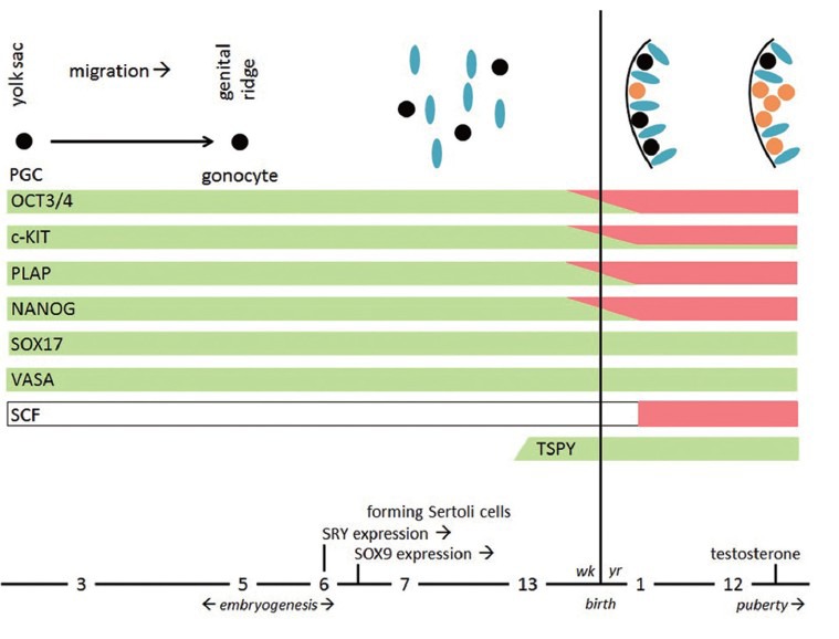
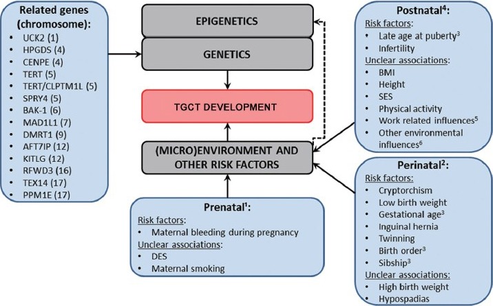
Malignant testicular germ cell tumors (TGCT) are the most frequent cancers in Caucasian males (20-40 years) with an 70% increasing incidence the last 20 years, probably due to combined action of (epi)genetic and (micro)environmental factors. It is expected that TGCT have carcinoma in situ(CIS) as their common precursor, originating from an embryonic germ cell blocked in its maturation process. The overall cure rate of TGCT is more than 90%, however, men surviving TGCT can present long-term side effects of systemic cancer treatment. In contrast, men diagnosed and treated for CIS only continue to live without these long-term side effects. Therefore, early detection of CIS has great health benefits, which will require an informative screening method. This review described the etiology and early pathogenesis of TGCT, as well as the possibilities of early detection and future potential of screening men at risk for TGCT. For screening, a well-defined risk profile based on both genetic and environmental risk factors is needed. Since 2009, several genome wide association studies (GWAS) have been published, reporting on single-nucleotide polymorphisms (SNPs) with significant associations in or near the genes KITLG, SPRY4, BAK1, DMRT1, TERT, ATF7IP, HPGDS, MAD1L1, RFWD3, TEX14, and PPM1E, likely to be related to TGCT development. Prenatal, perinatal, and postnatal environmental factors also influence the onset of CIS. A noninvasive early detection method for CIS would be highly beneficial in a clinical setting, for which specific miRNA detection in semen seems to be very promising. Further research is needed to develop a well-defined TGCT risk profile, based on gene-environment interactions, combined with noninvasive detection method for CIS.
Figures
References
-
- Friedman NB. Tumors of the testis. Am J Pathol. 1946;22:635–7. - PubMed
-
- Runyan C, Schaible K, Molyneaux K, Wang Z, Levin L, et al. Steel factor controls midline cell death of primordial germ cells and is essential for their normal proliferation and migration. Development. 2006;133:4861–9. - PubMed
-
- Donovan PJ. Growth factor regulation of mouse primordial germ cell development. Curr Top Dev Biol. 1994;29:189–225. - PubMed
-
- Wylie C. Germ cells. Cell. 1999;96:165–74. - PubMed
-
- Ulbright TM. Germ cell tumors of the gonads: a selective review emphasizing problems in differential diagnosis, newly appreciated, and controversial issues. Mod Pathol. 2005;18(Suppl 2):S61–79. - PubMed
Publication types
MeSH terms
Substances
LinkOut - more resources
Full Text Sources
Other Literature Sources
Medical
Miscellaneous
