. 2015 Oct;17(10):1374-85.
doi: 10.1093/neuonc/nov040. Epub 2015 Mar 19.

Melanoma brain metastasis is independent of lactate dehydrogenase A expression

Affiliations

Melanoma brain metastasis is independent of lactate dehydrogenase A expression

Terje Sundstrøm et al. Neuro Oncol. 2015 Oct.

Abstract

Background: The key metabolic enzyme lactate dehydrogenase A (LDHA) is overexpressed in many cancers, and several preclinical studies have shown encouraging results of targeted inhibition. However, the mechanistic importance of LDHA in melanoma is largely unknown and hitherto unexplored in brain metastasis.

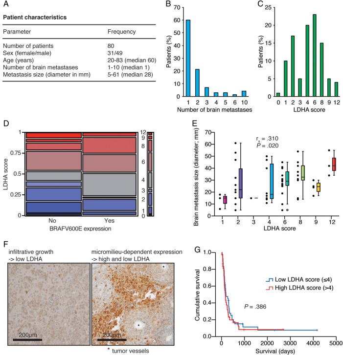
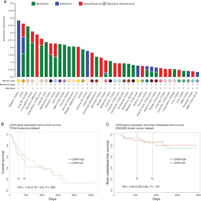
Methods: We investigated the spatial, temporal, and functional features of LDHA expression in melanoma brain metastasis across multiple in vitro assays, in a robust and predictive animal model employing MRI and PET imaging, and in a unique cohort of 80 operated patients. We further assessed the genomic and proteomic landscapes of LDHA in different cancers, particularly melanomas.

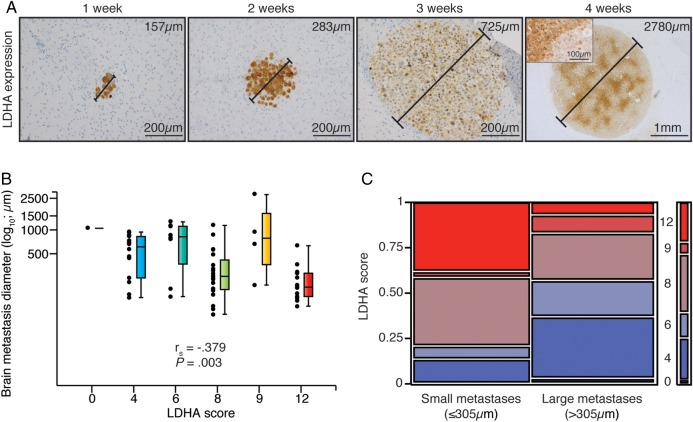
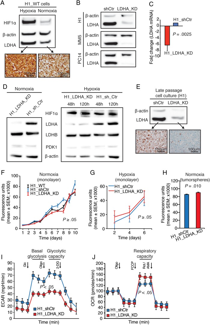
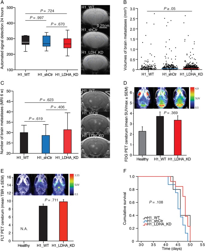
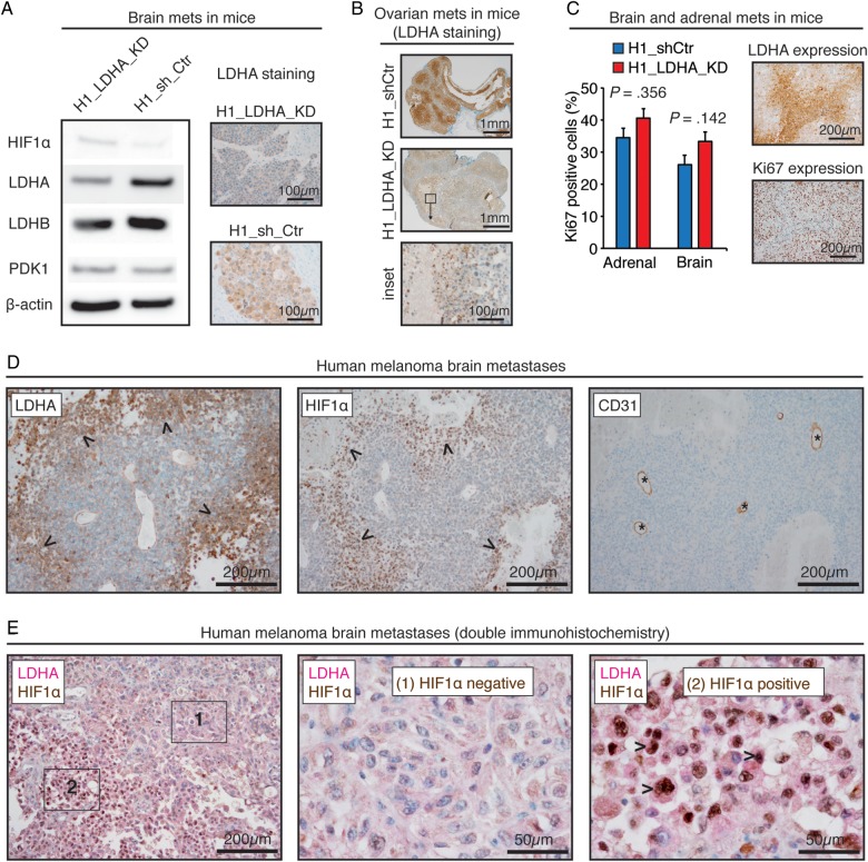
Results: LDHA expression was especially strong in early and small brain metastases in vivo and related to intratumoral hypoxia in late and large brain metastases in vivo and in patients. However, LDHA expression in human brain metastases was not associated with the number of tumors, BRAF(V600E) status, or survival. Moreover, LDHA depletion by small hairpin RNA interference did not affect cell proliferation or 3D tumorsphere growth in vitro or brain metastasis formation or survival in vivo. Integrated analyses of the genomic and proteomic landscapes of LDHA indicated that LDHA is present but not imperative for tumor progression within the CNS, or predictive of survival in melanoma patients.

Conclusions: In a large patient cohort and in a robust animal model, we show that although LDHA expression varies biphasically during melanoma brain metastasis formation, tumor progression and survival seem to be functionally independent of LDHA.

Keywords: LDHA; brain metastasis; lactate dehydrogenase A; melanoma; metabolism.


Figures

Fig. 1.
Fig. 1.
LDHA expression levels of melanoma brain metastases in mice displayed a biphasic pattern over time. (A) LDHA immunohistochemistry of brain metastases of increasing size (diameters provided) over 4 wk post intracardiac injection (n = 3 per wk). Inset displays representative staining pattern within the largest metastasis. (B) LDHA score vs brain metastasis size (n = 59; Spearman correlation). No metastases got an LDHA score 1–3. (C) LDHA scores of small (≤305 μm; n = 30) vs large (>305 μm; n = 29) brain metastases (median split; Pearson chi-square test, P = .038).
Fig. 2.
Fig. 2.
LDHA expression levels of human melanoma brain metastases was not predictive of tumor burden or survival but increased with tumor size. (A) Cohort characteristics. (B) Number of brain metastases. (C) LDHA scores. (D) BRAFV600E expression status vs LDHA score (Pearson chi-square test, P = .221). (E) LDHA score vs brain metastasis size (n = 56; Spearman correlation). (F) LDHA immunohistochemistry of diffusely infiltrating tumor cells (left) and micromilieu-dependent expression in the tumor core (right). (G) Kaplan–Meier survival plot of low vs high LDHA score (median split; Mantel–Cox log-rank test). Five patients in the low group and 7 patients in the high group were still alive or lost to follow-up.
Fig. 3.
Fig. 3.
Cellular LDHA knockdown was effective and stable but did not affect cellular proliferation or tumorsphere growth. (A) Western blots of H1_WT cells in hypoxia and normoxia (48 h) with corresponding LDHA immunocytochemistry. (B) Western blots of control (sh_Ctr) and LDHA knockdown (LDHA_KD) derivatives of H1 and 2 other human metastasis cell lines (MELMET5 [MM5]/melanoma and PC14-PE6_Br2 [PC14]/lung cancer). (C) LDHA mRNA levels in H1 derivatives. Mean knockdown efficiency was 83 ± 3% (n = 3; mean ± SEM; Student t-test). (D) Western blots of H1_LDHA_KD and H1_shCtr cells in normoxia (48 h) and acute (48 h) and chronic (120 h) hypoxia (second passage after green fluorescent protein–positive fluorescence activated cell sorting). (E) Western blot of cells cultured continuously for 2 months with corresponding LDHA immunocytochemistry. (F) Proliferation assay in normoxia (n = 6 per cell line per time point; Kruskal–Wallis test). (G) Proliferation assay in hypoxia (n = 12 per cell line per time point; Student t-test). (H) Tumorsphere assay in normoxia (n = 36 per cell line; Student t-test). (I) Glycolytic stress test in normoxia (n = 3 per cell line per time point; mean ± SEM; Student t-test). (J) Mito stress test in normoxia (n = 2 per cell line per time point; mean ± SEM; Student t-test). (A, D, and G) Hypoxia (1% O2).
Fig. 4.
Fig. 4.
LDHA knockdown did not affect brain metastasis or survival in mice. (A) MRI-based automated quantification of nanoparticle-labeled melanoma cells in mouse brains 24 h post intracardiac injection of 5 × 10⁵ cells (Student t-test). Typical brain MRI T2*-weighted images with an overlay of detected signals. (B) Volumes of individual brain metastases at the 6-wk MRI (Mann–Whitney U test, H1_WT vs H1_shCtr, P = .413; H1_WT vs H1_LDHA_KD, P = .715; H1_LDHA_KD vs H1_shCtr, P = .475). (C) Number of brain metastases at the 6-wk MRI (T1-weighted images with contrast; Student t-test). (D) Maximum standardized uptake value (SUVmax) on 18F-FDG PET cerebrum at 6 wk (n = 3 per group; Student t-test). Note the high signal intensity in the frontal (Harderian glands) and cerebellar regions in healthy control mice in the combined PET and CT maximum intensity projection (MIP) images. (E) Tumor-to-brain ratio (TBR; SUVmax tumor/SUVmax normal brain) on 18F-FLT PET cerebrum at 6 wk (combined PET and CT MIP images; n = 2 per group; Student t-test). Note the lack of signals in healthy mice, as 18F-FLT does not cross an intact blood–brain barrier (hence, not applicable). (F) Kaplan–Meier survival plot (Mantel–Cox log-rank test). (A–C and F) H1_WT group, n = 5; H1_shCtr group, n = 8; and H1_LDHA_KD group, n = 13.
Fig. 5.
Fig. 5.
LDHA knockdown was stable in vivo and hypoxia dependent in human melanoma brain metastases. (A) Western blots of brain metastases (mets) in mice with corresponding LDHA immunohistochemistry. (B) LDHA immunohistochemistry of ovarian metastases in mice with inset showing perinecrotic regulation. (C) Ki67-positive cells in brain and adrenal metastases in mice (n = 5; mean ± SEM; Student t-test) with adjacent images showing regionally distinct Ki67 and LDHA patterns. (D) Immunohistochemistry of a human melanoma brain metastasis displaying a spatial overlap of LDHA and HIF1α expression (arrowheads) ∼150 μm from CD31-positive vessels (asterisks). (B) Double immunohistochemistry of a human melanoma brain metastasis and 2 insets showing spatial coexpression of perinuclear LDHA and nuclear HIF1α (arrowheads).
Fig. 6.
Fig. 6.
Genomic alterations of LDHA were infrequent in human melanomas and other cancers, and LDHA expression was not predictive of survival. (A) Histogram showing a summary of LDHA mutations, deletions, and amplifications across 40 of the 69 studies from the cBioPortal (19 studies did not report any alterations). Three melanoma series are featured here: TCGA (provisional), Broad, and Yale. Abbreviations: CNA (copy number alterations), CCLE (Cancer Cell Line Encyclopedia), SC (small cell), adeno (adenocarcinoma), CLCGP (Clinical Lung Cancer Genome Project), MSKCC (Memorial Sloan Kettering Cancer Center), JHU (Johns Hopkins University), ACC (adrenocortical carcinoma), GBM (glioblastoma multiforme), NCI-60 (National Cancer Institute panel of 60 cell lines), CS (carcinosarcoma), ACyC (adenoid cystic carcinoma), Squ (squamous cell carcinoma), and AMC (Asan Medical Center). (B) Overall survival between low and high LDHA expression groups in 163 TCGA melanoma patients. (C) Brain metastasis–free survival in 82 breast cancer patients from the GSE2603 dataset. (B and C) Cohorts were divided at median of gene expression. Cox proportional hazard ratios (HR) (with CIs) are provided with log-rank test P-values.

Similar articles

Cited by

References

    1. MacKie RM, Hauschild A, Eggermont AM. Epidemiology of invasive cutaneous melanoma. Ann Oncol. 2009;20(suppl. 6):vi1–vi7. - PMC - PubMed
    1. Eggermont AM, Spatz A, Robert C. Cutaneous melanoma. Lancet. 2014;383(9919):816–827. - PubMed
    1. Flanigan JC, Jilaveanu LB, Chiang VL, et al. Advances in therapy for melanoma brain metastases. Clin Dermatol. 2013;31(3):264–281. - PubMed
    1. Vander Heiden MG, Cantley LC, Thompson CB. Understanding the Warburg effect: the metabolic requirements of cell proliferation. Science. 2009;324(5930):1029–1033. - PMC - PubMed
    1. Warburg O. On the origin of cancer cells. Science. 1956;123(3191):309–314. - PubMed

Publication types