Review
. 2015 Apr;14(4):420-34.
doi: 10.1016/S1474-4422(14)70201-7.

Congenital myasthenic syndromes: pathogenesis, diagnosis, and treatment

Affiliations
Review

Congenital myasthenic syndromes: pathogenesis, diagnosis, and treatment

Andrew G Engel et al. Lancet Neurol. 2015 Apr.

Erratum in

Abstract

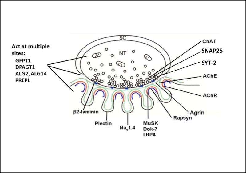
The congenital myasthenic syndromes (CMS) are a diverse group of genetic disorders caused by abnormal signal transmission at the motor endplate, a special synaptic contact between motor axons and each skeletal muscle fibre. Most CMS stem from molecular defects in the muscle nicotinic acetylcholine receptor, but they can also be caused by mutations in presynaptic proteins, mutations in proteins associated with the synaptic basal lamina, defects in endplate development and maintenance, or defects in protein glycosylation. The specific diagnosis of some CMS can sometimes be reached by phenotypic clues pointing to the mutated gene. In the absence of such clues, exome sequencing is a useful technique for finding the disease gene. Greater understanding of the mechanisms of CMS have been obtained from structural and electrophysiological studies of the endplate, and from biochemical studies. Present therapies for the CMS include cholinergic agonists, long-lived open-channel blockers of the acetylcholine receptor ion channel, and adrenergic agonists. Although most CMS are treatable, caution should be exercised as some drugs that are beneficial in one syndrome can be detrimental in another.


Conflict of interest statement

Conflicts of Interest

None of the authors reports a conflict of interest.

Figures

Figure 1
Figure 1
Schematic diagram of an EP with locations of pre-synaptic, synaptic and postsynaptic CMS disease proteins. Green line, synaptic basal lamina; red line, AChR on crests of the junctional folds; blue line, LRP4, MuSK, Dok-7, and rapsyn closely associated with AChR. SC, Schwann cell; NT, nerve terminal. (Modified from figure 1, Neuromuscul Disord 2012;22:99–111).
Figure 2
Figure 2
ChAT deficiency. (A) Subtetanic stimulation rapidly decreases the endplate potential (EPP) which returns to the baseline slowly over more than 10 min. 3,4-diaminopyridine (3,4-DAP) which increases quantal release accelerates the decline of the EPP, whereas a low Ca2+/high Mg2+ solution which reduces quantal release prevents the abnormal decline of the EPP. (B) Positions of mutated residues in active site tunnel of ChAT, kinetic landscapes of wild-type and mutant enzymes obtained from reaction velocities over a range and AcCoA concentrations, and normalized kinetic parameters of wild-type and mutant ChAT. The catalytic efficiencies of the enzymes with AcCoA (kcat/Kma), choline (kcat/Kmb), and both substrates (kcat/KiaKmb), were calculated as described in Reference 11. The catalytic efficiencies of the mutant enzymes are <0.1% of wild-type. Kcat = maximal reaction velocity/enzyme concentration; KmA and KmB = Michelis-Menten constants for AcCoA and choline; Kia = dissociation constant for the enzyme-AcCoA complex. (Panel A is reproduced from Nature Rev Neurosci, 2003;330–352; Panel B is reproduced from Hum Mutat 2011;32:1259–1267.)
Figure 3
Figure 3
EP AChE deficiency. (A) Schematic diagram showing domains of a ColQ strand with 24 identified ColQ mutations. (B) Schematic diagram of the A12 species of asymmetric AChE. PRAD = proline rich attachment domain; HSPBD, heparan sulfate proteoglycan binding domain. (C) Electron cytochemical localization of AChE in a control subject (C) and in an AChE-deficient patient (D). There is no reaction for AChE at the patient EP. Bars in (C) and (D) = 1 μm. (Panel A is reproduced by from Neuromuscul Disord 22:99–111, 2012)
Figure 4
Figure 4
EP AChR deficiency. (A) Schematic diagram of low-expressor and null mutations reported in the AChR α, β, δ, and ɛ subunits. Most mutations occur in the ɛ subunit. Splice-site mutations point to the N-terminal codons of the predicted skipped exons. Panels (B) and (C) show ultrastructural localization of AChR with peroxidase-labeled α-bungartoxin at an EP from a control subject and from a patient homozygous for c.ɛ553del7. Bars in (B) and (C) = 1 μm. (Panel A is reproduced from Muscle Nerve, 27:4–25, 2003)
Figure 5
Figure 5
Scaled linear model of MUSK with its domains and observed mutations. Exon 2–3 skip predicts in-frame deletion. Red boxes in the frameshifting mutations represent missense amino acids. Transmembrane domain includes amino acids 496–516. FZ, frizzled; TM, transmembrane.
Figure 6
Figure 6
Degenerating EP in Dok7 myasthenia. Note replacement of degenerate junctional folds by debris, and large myeloid structure in junctional sarcoplasm. Bar = 1 μm.
Figure 7
Figure 7
CMS due to mutations in rapsyn. (A) Rapsyn domains and interactions. (B) Small cholinesterase reactive EP regions are distributed over an extended length of the muscle fiber. (C) and (D) Multiple small nerve terminals appear over poorly developed junctional folds. In (C), the distribution of AChR on the postsynaptic membrane, visualized with peroxidase-labeled α-bungarotoxin, is patchy. Bars 50 μm in (A) and 1 μm in (B) and (C).(Reproduced from Neurology 2009;73:228–235.)

Similar articles

Cited by

References

    1. Wood SJ, Slater CP. Safety factor at the neuromuscular junction. Prog Neurobiol. 2001;64:393–429. - PubMed
    1. Tsujino A, Maertens C, Ohno K, et al. Myasthenic syndrome caused by mutation of the SCN4A sodium channel. Proc Natl Acad Sci USA. 2003;100:7377–7382. - PMC - PubMed
    1. Abicht A, Dusl M, Guergueltcheva V, et al. Congenital myasthenic syndromes: achievements and limitations of phenotype-guided gene-after-gene sequencing in diagnostic practice: a study of 680 patients. Hum Mutat. 2014;33:1474–1484. - PubMed
    1. Senderek J, Muller JS, Dusl M, et al. Hexosamine biosynthetic pathway mutations cause neuromuscular transmission defect. Am J Hum Genet. 2011;88:162–172. - PMC - PubMed
    1. Shinawi M, Cheung SW. The array CGH and its clinical applications. Drug Discov Today. 2008;13:760–770. - PubMed

Publication types

MeSH terms