. 2015;7(5):530-44.
doi: 10.1159/000375161. Epub 2015 Mar 19.

Antiviral and Immunoregulatory Effects of Indoleamine-2,3-Dioxygenase in Hepatitis C Virus Infection

Affiliations

Antiviral and Immunoregulatory Effects of Indoleamine-2,3-Dioxygenase in Hepatitis C Virus Infection

Quentin Lepiller et al. J Innate Immun. 2015.

Abstract

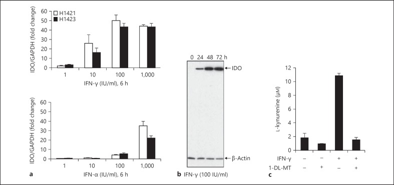
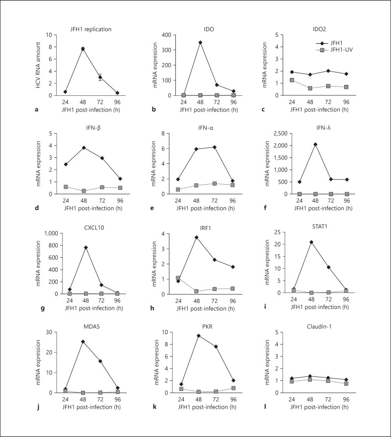
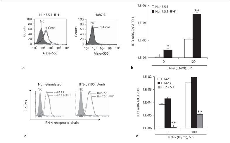
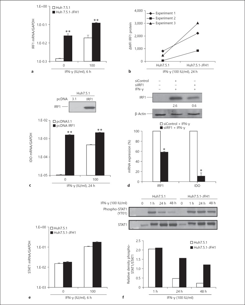
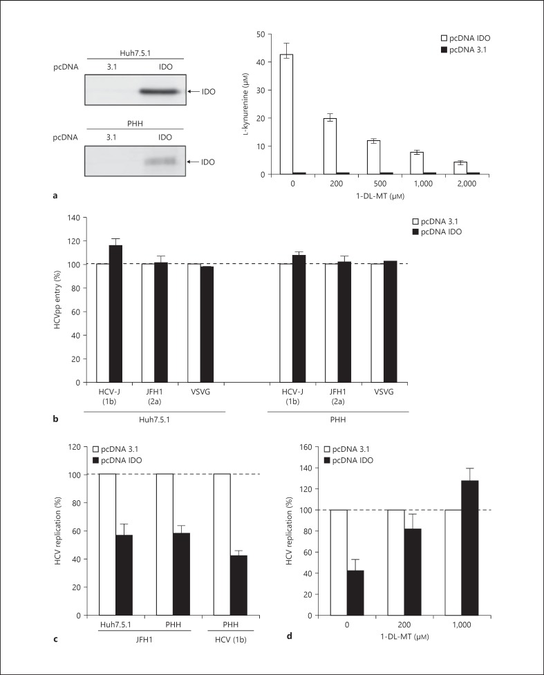
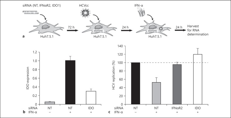
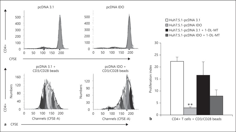
In patients with hepatitis C virus (HCV) infection, enhanced activity of indoleamine-2,3-dioxygenase 1 (IDO) has been reported. IDO - a tryptophan-catabolizing enzyme - has been considered as both an innate defence mechanism and an important regulator of the immune response. The molecular mechanism of IDO induction in HCV infection and its role in the antiviral immune response remain unknown. Using primary human hepatocytes, we show that HCV infection stimulates IDO expression. IDO gene induction was transient and coincided with the expression of types I and III interferons (IFNs) and IFN-stimulated genes in HCV-infected hepatocytes. Overexpression of hepatic IDO prior to HCV infection markedly impaired HCV replication in hepatocytes, suggesting that IDO limits the spread of HCV within the liver. siRNA-mediated IDO knock-down revealed that IDO functions as an IFN-mediated anti-HCV effector. Hepatic IDO was most potently induced by IFN-x03B3;, and ongoing HCV replication could significantly upregulate IDO expression. IRF1 (IFN-regulatory factor 1) and STAT1 (signal transducer and activator of transcription 1) regulated hepatic IDO expression. Hepatic IDO expression also had a significant inhibitory effect on CD4+ T-cell proliferation. Our data suggest that hepatic IDO plays a dual role during HCV infection by slowing down viral replication and also regulating host immune responses.


Figures

Fig. 1
Fig. 1
Expression of IDO in PHH. a PHH from 2 donors (H1421, H1423) were incubated with different doses of IFNs. IDO expression is shown as a ratio of IDO/GAPDH compared to non-stimulated PHH. b PHH were incubated with IFN-γ and harvested at the indicated time points. Levels of IDO protein expression in PHH lysates were determined by Western blotting. β-Actin levels are shown as a loading control. c PHH were stimulated with IFN-γ (100 IU/ml) for 48 h in the presence and absence of 1-DL-MT (100 µM). Then, cells were cultured for 4 h in HBSS buffer containing 100 µM of tryptophan. Kynurenine accumulation was measured in cell culture supernatants by HPLC. Data are given as mean kynurenine production ± SD from 2 independent experiments.
Fig. 2
Fig. 2
HCV infection stimulates IDO expression in PHH. a PHH (donor H0199) were infected with JFH1 for 3 h, washed and then lysed at the time points indicated. HCV RNA levels were normalized to GAPDH mRNA levels. Results are presented as fold induction to 3-hour post-infected PHH. Gene induction was measured by real-time RT-PCR analysis in PHH exposed to JFH1 or UV-infected JFH1. Gene induction of IDO (b), IDO2 (c), IFN-β (d), IFN-α (e), IFN-λ (f), CXCL10 (g), IRF1 (h), STAT1 (i), MDA5 (j), PKR (k), and claudin-1 (l) are shown as fold change over uninfected PHH cultures.
Fig. 3
Fig. 3
Ongoing HCV replication in Huh7.5.1 cells enhances IFN-γ-induced IDO expression. NC = Negative control. a Robust HCV infection of Huh7.5.1 cells was determined using anti-HCV core staining and flow cytometry at day 5 after inoculation. b JFH1-infected Huh7.5.1 cells and uninfected control cells were stimulated with IFN-γ and then analysed for IDO gene expression. c After 4 h of IFN-γ stimulation, JFH1-infected Huh7.5.1 cells and control cells were analysed for IFN-γ receptor α-chain expression. d IDO gene expression levels in PHH (donor H1421, H1423) and Huh7.5.1 cells in the presence or absence of IFN-γ. * p < 0.05, ** p < 0.01.
Fig. 4
Fig. 4
IRF1 regulates IDO expression in HCV infection. MFI = Mean fluorescence intensity. a JFH1-infected Huh7.5.1 cells and uninfected control cells were analysed for IRF1 expression in the absence and presence of IFN-γ. IRF1 expression is shown as a ratio of IRF1/GAPDH. b IFN-γ-induced IRF1 protein expression is shown as delta mean fluorescence intensity (ΔMFI: MFI of IFN-γ-stimulated cells minus MFI of non-stimulated cells). c Huh7.5.1 cells were transfected with pcDNA IRF1 or pcDNA3.1. IRF1 expression was verified by Western blotting 48 h after transfection. IDO mRNA expression is shown in the presence or absence of IFN-γ in transfected cells. d IRF1 was silenced in Huh7.5.1 cells. Numbers represent densitometric analysis of IRF1 bands protein normalized to β-actin levels. Then, cells were stimulated with IFN-γ (50 IU/ml) for 7 h, and the relative levels of IRF1 and IDO mRNA expression were determined. Data are expressed as the percentage of gene expression for IRF1-silenced Huh7.5.1 cells in comparison to siControl (= 100%). e JFH1-infected Huh7.5.1 cells and control cells were analysed for STAT1 expression in the absence and presence of IFN-γ. STAT1 expression is shown as a ratio of STAT1/GAPDH. f After treatment with IFN-γ, cells were harvested at the indicated times. Equal amounts of total cell lysates were immunoblotted with anti-STAT1 and anti-phospho-STAT1 antibodies. Densitometry analysis shows phospho-STAT1 levels relative to STAT1 levels. * p < 0.05, ** p < 0.01.
Fig. 5
Fig. 5
Impact of hepatic IDO expression on HCV infection. a Huh7.5.1 cells and PHH (H0204, H0216, H0208) were transfected with pcDNA IDO or pcDNA3.1. IDO protein expression was verified by Western blotting. Functional activity of the expressed IDO protein in Huh7.5.1 cells was verified by the accumulation of kynurenine in the supernatant in the presence and absence of the specific IDO inhibitor 1-DL-MT using the Ehrlich reagent method. b At 48 h after transfection, Huh7.5.1 cells and PHH (donor H0204, H0216, H0208) were assessed for HCVpp entry (genotype 2a: JFH1, genotype 1b: HCV-J). VSVG = Vesicular stomatitis virus G glycoprotein. Data are expressed as the percentage of HCVpp entry into IDO-transfected PHH compared to pcDNA3.1-transfected control cells (= 100%). Vesicular stomatitis virus G glycoprotein-pseudotyped virions were included as a control. c At 48 h after transfection, Huh7.5.1 cells and PHH were incubated with JFH1 or HCV serum (genotype 1b). At 2 days after JFH1 infection, viral replication in IDO-transfected cells was measured. HCV RNA copies were normalized to GAPDH. Data are shown as mean percentage of HCV replication compared to pcDNA3.1-transfected cells (= 100%). d Rescued HCV JFH1 replication in IDO-transfected Huh7.5.1 cells. JFH1 replication in IDO-transfected Huh7.5.1 cells was determined in the presence and absence of the specific IDO inhibitor 1-DL-MT. Data are shown as the mean ± SD of triplicate determinations and are representative of 3 independent experiments.
Fig. 6
Fig. 6
IDO function as an ISG effector with anti-HCV activity. NT = Non-targeting control. a Flowchart describing the 3 main parts of the experiment. Huh7.5.1 cells were transfected with siRNAs against IFNαR2, IDO or NT siRNAs. After 72 h, cells were infected with HCV JFH1 virus and, after an additional 24 h, IFN-α (10 IU/ml) was added. Cells were harvested 24 h later, and HCV replication was measured by qRT-PCR. b Knock-down efficiencies of IDO. IDO mRNA levels were quantified by qRT-PCR at day 5 after siRNA transfection. c Intracellular HCV RNA amounts. Data are shown as mean percentage of HCV replication compared to cells treated with non-targeting control siRNAs (= 100%). Values represent the mean ± SD of triplicate measurements.
Fig. 7
Fig. 7
Hepatic IDO expression inhibits CD4+ T cell proliferation. a Flow cytometry analysis of CD4 and CFSE staining following CD4+T/Huh7.5.1 cell co-culture. Huh7.5.1 cells were transfected with pcDNA IDO or pcDNA3.1. At 48 h after transfection, cells were added to CD3/CD28-activated CD4+ T cells or control CD4+ T cells. CFSE fluorescence of undivided non-stimulated and CD3/CD28-activated CD4+ T cells undergoing multiple rounds of cell division following Huh7.5.1 co-culture is shown. b Pooled data from 3 different co-culture experiments are shown as mean proliferation index ± SD. Data were compiled from 3 different healthy CD4+ T-cell donors. Inhibition of CD4+ T-cell proliferation by IDO-transfected Huh7.5.1 cells was statistically significant. ** p < 0.01. The presence of 1-DL-MT (1,000 µM) for the duration of the co-culture partially restored IDO-mediated CD4+ T-cell suppression.

Similar articles

Cited by

References

    1. Liang TJ, Heller T. Pathogenesis of hepatitis C-associated hepatocellular carcinoma. Gastroenterology. 2004;127:S62–S71. - PubMed
    1. Horner SM, Gale M. Regulation of hepatic innate immunity by hepatitis C virus. Nat Med. 2013;19:879–888. - PMC - PubMed
    1. Rehermann B. Pathogenesis of chronic viral hepatitis: differential roles of T cells and NK cells. Nat Med. 2013;19:859–868. - PMC - PubMed
    1. Mellor AL, Munn DH. IDO expression by dendritic cells: tolerance and tryptophan catabolism. Nat Rev Immunol. 2004;4:762–774. - PubMed
    1. Pfefferkorn ER. Interferon gamma blocks the growth of Toxoplasma gondii in human fibroblasts by inducing the host cells to degrade tryptophan. Proc Natl Acad Sci U S A. 1984;81:908–912. - PMC - PubMed

Publication types

MeSH terms