Review
. 2015 Jun;129(6):775-88.
doi: 10.1007/s00401-015-1410-7. Epub 2015 Mar 20.

Pilocytic astrocytoma: pathology, molecular mechanisms and markers

Affiliations
Review

Pilocytic astrocytoma: pathology, molecular mechanisms and markers

V Peter Collins et al. Acta Neuropathol. 2015 Jun.

Abstract

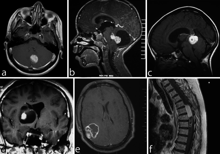
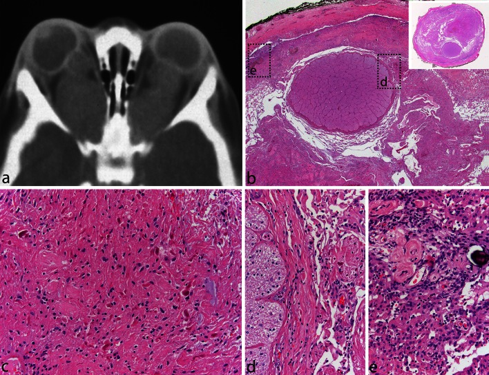
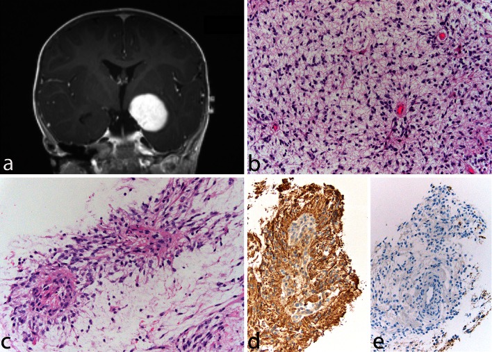
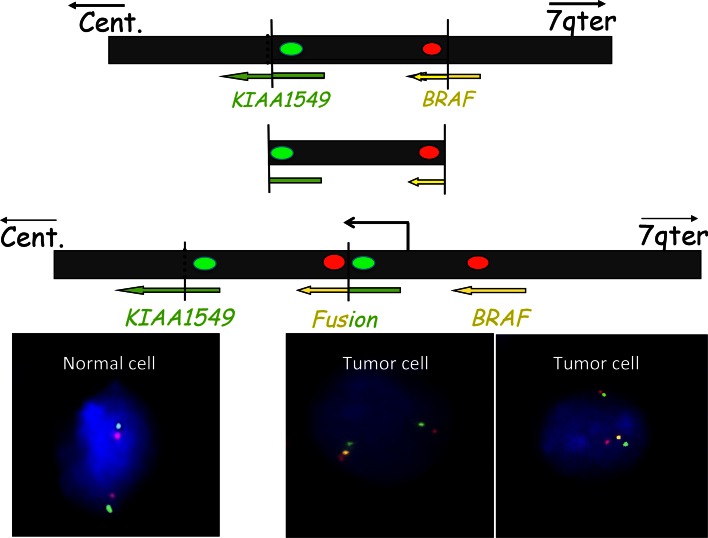
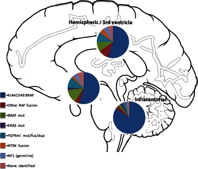
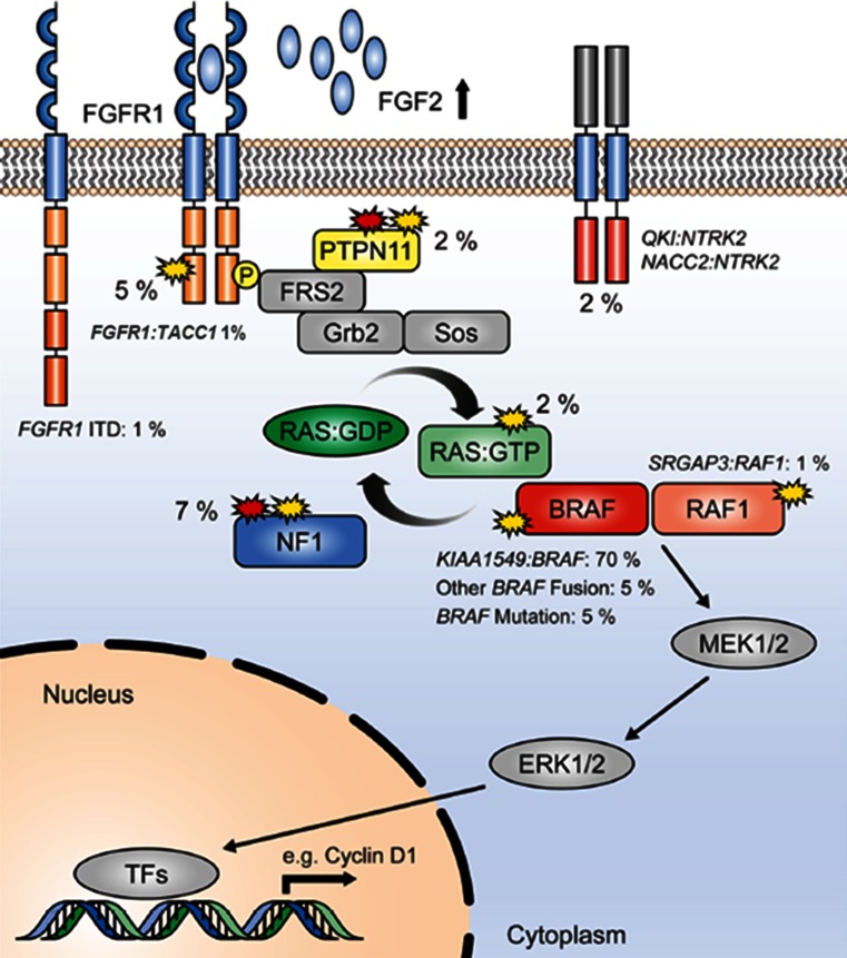
Pilocytic astrocytomas (PAs) were recognized as a discrete clinical entity over 70 years ago. They are relatively benign (WHO grade I) and have, as a group, a 10-year survival of over 90%. Many require merely surgical removal and only very infrequently do they progress to more malignant gliomas. While most show classical morphology, they may present a spectrum of morphological patterns, and there are difficult cases that show similarities to other gliomas, some of which are malignant and require aggressive treatment. Until recently, almost nothing was known about the molecular mechanisms involved in their development. The use of high-throughput sequencing techniques interrogating the whole genome has shown that single abnormalities of the mitogen-activating protein kinase (MAPK) pathway are exclusively found in almost all cases, indicating that PA represents a one-pathway disease. The most common mechanism is a tandem duplication of a ≈2 Mb-fragment of #7q, giving rise to a fusion between two genes, resulting in a transforming fusion protein, consisting of the N-terminus of KIAA1549 and the kinase domain of BRAF. Additional infrequent fusion partners have been identified, along with other abnormalities of the MAP-K pathway, affecting tyrosine kinase growth factor receptors at the cell surface (e.g., FGFR1) as well as BRAF V600E, KRAS, and NF1 mutations among others. However, while the KIAA1549-BRAF fusion occurs in all areas, the incidence of the various other mutations identified differs in PAs that develop in different regions of the brain. Unfortunately, from a diagnostic standpoint, almost all mutations found have been reported in other brain tumor types, although some retain considerable utility. These molecular abnormalities will be reviewed, and the difficulties in their potential use in supporting a diagnosis of PA, when the histopathological findings are equivocal or in the choice of individualized therapy, will be discussed.


Figures

Fig. 1
Fig. 1
Pilocytic astrocytoma, with its characteristic imaging features, may occur virtually at any site in the CNS. Six different examples (all histologically confirmed) with strong contrast enhancement are illustrated: two cerebellar examples, one of a small left para-vermian well-circumscribed and solid tumor (a) and one of a cystic tumor with a mural nodule (b); a “dorsally exophytic” midbrain PA (c); a cyst with a mural nodule occupying the right thalamus (d); a peripheral solid and cystic tumor in the right parietal lobe (e); and a large, circumscribed, intramedullary, tumor with a cystic component (f)
Fig. 2
Fig. 2
Pilocytic astrocytoma of the optic nerve. Bilateral fusiform enlargement of the optic nerve is virtually diagnostic of neurofibromatosis type 1 (a). The tumor typically extends into the leptomeningeal space, expanding the dural sheath and compressing the remaining optic nerve proper, which is atrophic (b). A complete cross section of the optic nerve is shown in the inset. The tumor has classic PA features with a densely fibrillated appearance and numerous Rosenthal fibers (c). The interface between the optic nerve and the tumor is shown in (d), while the subdural region shows meningothelial hyperplasia, at times, with scant psammoma bodies (e)
Fig. 3
Fig. 3
Pilomyxoid astrocytoma. A 13-month-old boy presented with a relatively circumscribed, strongly enhancing left medial temporal lobe mass (a). The tumor shows a monomorphous cell population in a loosely arranged myxoid background (b). Tumor cells with angiocentric arrangement and formation of pseudopapillary structures is typical (c). GFAP is typically positive in tumor cells (d), and immunocytochemistry for neurofilaments is negative, the tumor being generally relatively solid and devoid of axons (e)
Fig. 4
Fig. 4
The common fusion rearrangement: The upper black box represents 7q34 on the long arm of chromosome 7. Both KIAA1549 and BRAF read towards the centromere (cent). A fragment of approximately 2 MB is duplicated and inserted at the breakpoint, producing a tandem duplication and the fusion between the 5′ end of KIAA1549 and the 3′ end of the BRAF gene that codes for the kinase domain. The fusion gene thus codes for the BRAF kinase domain together with the N-terminal part of KIAA1549, replacing the BRAF regulatory domain. It is important to remember that the exact breakpoints vary, resulting in nine combinations of KIAA1549-BRAF exons, all with an open reading frame from KIAA1549 spliced sequence into the BRAF sequence. This makes simple RT-PCR assays difficult. The red and green dots represent the location of FISH probes that could be used to identify the occurrence of the tandem duplication as demonstrated in the lower part of the figure, showing interphase normal and tumor nuclei with the tandem duplication hybridized with such probes. Note that the two unraveled normal chromosomes 7 in the normal nucleus show a single red and green signal adjacent to each other, while the tumor cell nuclei show one normal chromosomal signal but also a signal from the second chromosome 7, showing, in addition, a yellow signal (due to the fusion of the extra, now adjacent, red and green signals)
Fig. 5
Fig. 5
Pie charts summarizing the estimated frequency of particular MAPK pathway alterations in different anatomic locations (posterior fossa, diencephalon and cerebral hemispheres), calculated from a total of 188 PAs described in described in Zhang et al. [67] and Jones et al. [32]
Fig. 6
Fig. 6
Summary showing the MAP kinase pathway with the approximate incidence of the different mutations in percent in a series of PAs (adapted with permission from Jones et al. [32])
Fig. 7
Fig. 7
Pilocytic astrocytoma and rosette-forming glioneuronal tumor of the fourth ventricle. This solid and cystic tumor, occurring in a 26-year-old man, occupied the superior portion of the fourth ventricle (a). Most of the tumor displayed classic features of PA, being biphasic with microcystic areas (b) and areas with densely fibrillated tumors cells with abundant Rosenthal fibers (c). A small, distinct, very soft and light gray component was noted grossly, corresponding histologically to a classic “rosette-forming glioneuronal component” (d), with its characteristic high-power appearance (e) and synaptophysin positivity, corresponding to the center of the neuropil rosettes (f)

References

    1. Alpers CE, Davis RL, Wilson CB. Persistence and late malignant transformation of childhood cerebellar astrocytoma. Case report. J Neurosurg. 1982;57:548–551. doi: 10.3171/jns.1982.57.4.0548. - DOI - PubMed
    1. Antonelli M, Badiali M, Moi L, Buttarelli FR, Baldi C, Massimino M, et al. KIAA1549:BRAF fusion gene in pediatric brain tumors of various histogenesis. Pediatr Blood Cancer. 2014 - PubMed
    1. Aoki Y, Niihori T, Narumi Y, Kure S, Matsubara Y. The RAS/MAPK syndromes: novel roles of the RAS pathway in human genetic disorders. Hum Mutat. 2008;29:992–1006. doi: 10.1002/humu.20748. - DOI - PubMed
    1. Badiali M, Gleize V, Paris S, Moi L, Elhouadani S, Arcella A, et al. KIAA1549-BRAF fusions and IDH mutations can coexist in diffuse gliomas of adults. Brain Pathol. 2012;22:841–847. doi: 10.1111/j.1750-3639.2012.00603.x. - DOI - PMC - PubMed
    1. Bar EE, Lin A, Tihan T, Burger PC, Eberhart CG. Frequent gains at chromosome 7q34 involving BRAF in pilocytic astrocytoma. J Neuropathol Exp Neurol. 2008;67:878–887. doi: 10.1097/NEN.0b013e3181845622. - DOI - PubMed

Publication types

MeSH terms

LinkOut - more resources