

Monocytes in myocardial infarction
- PMID: 25792449
- PMCID: PMC4409536
- DOI: 10.1161/ATVBAHA.114.304652
Monocytes in myocardial infarction
Abstract
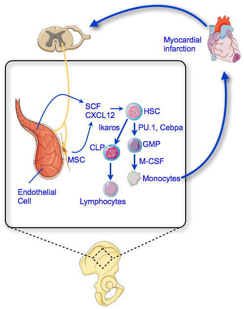
Myocardial infarction (MI) is the leading cause of death in developed countries. Though timely revascularization of the ischemic myocardium and current standard therapy reduce acute mortality after MI, long-term morbidity and mortality remain high. During the first 1 to 2 weeks after MI, tissues in the infarcted myocardium undergo rapid turnover, including digestion of extracellular matrix and fibrosis. Post-MI repair is crucial to survival. Monocytes recruited to the infarcted myocardium remove debris and facilitate the repair process. However, exaggerated inflammation may also impede healing, as demonstrated by the association between elevated white blood cell count and in-hospital mortality after MI. Monocytes produced in the bone marrow and spleen enter the blood after MI and are recruited to the injured myocardium in 2 phases. The first phase is dominated by Ly-6c(high) monocytes and the second phase by Ly-6c(low) monocytes. Yet the number of Ly6C(low) monocytes recruited to the infarct is much lower, and Ly6C(high) monocytes can differentiate to Ly6C(low) macrophages in later healing stages. Understanding the signals regulating monocytosis after MI will help design new therapies to facilitate cardiac healing and limit heart failure.
Keywords: hematopoiesis; macrophages; monocytes; myocardial infarction.
© 2015 American Heart Association, Inc.
Figures
References
-
- Auffray C, Sieweke MH, Geissmann F. Blood monocytes: development, heterogeneity, and relationship with dendritic cells. Annu Rev Immunol. 2009;27:669–692. - PubMed
-
- Auffray C, Fogg D, Garfa M, Elain G, Join-Lambert O, Kayal S, Sarnacki S, Cumano A, Lauvau G, Geissmann F. Monitoring of blood vessels and tissues by a population of monocytes with patrolling behavior. Science. 2007;317:666–670. - PubMed
-
- Sieweke MH, Allen JE. Beyond stem cells: self-renewal of differentiated macrophages. Science. 2013;342:1242974. - PubMed
Publication types
MeSH terms
Grants and funding
LinkOut - more resources
Full Text Sources
Medical
