. 2015 Mar 15;29(6):646-57.
doi: 10.1101/gad.258731.115.

A retrovirus packages nascent host noncoding RNAs from a novel surveillance pathway

Affiliations

A retrovirus packages nascent host noncoding RNAs from a novel surveillance pathway

Matthew J Eckwahl et al. Genes Dev. .

Abstract

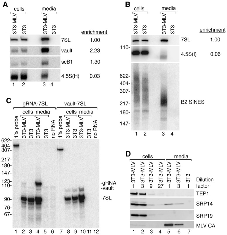
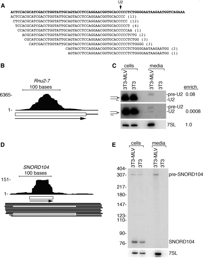
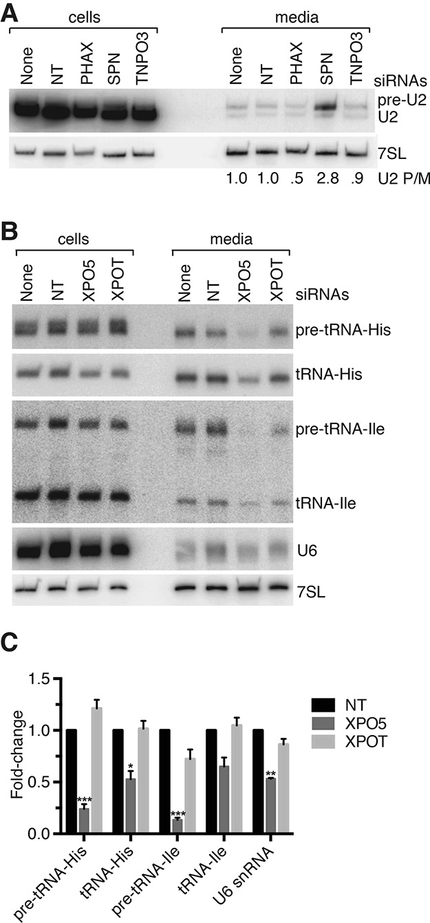
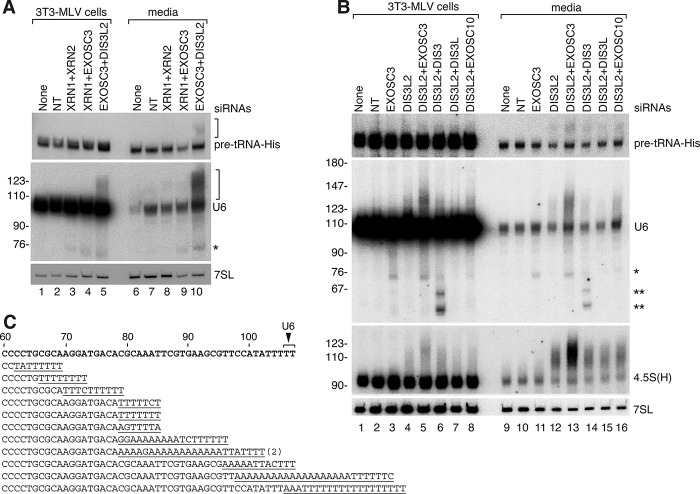
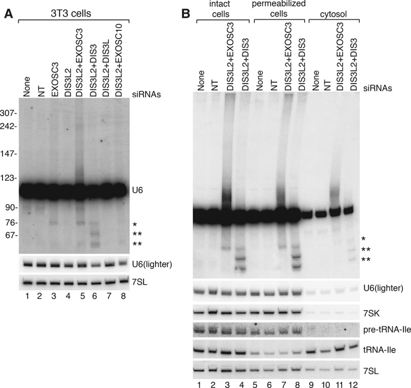
Although all retroviruses recruit host cell RNAs into virions, both the spectrum of RNAs encapsidated and the mechanisms by which they are recruited remain largely unknown. Here, we used high-throughput sequencing to obtain a comprehensive description of the RNAs packaged by a model retrovirus, murine leukemia virus. The major encapsidated host RNAs are noncoding RNAs (ncRNAs) and members of the VL30 class of endogenous retroviruses. Remarkably, although Moloney leukemia virus (MLV) assembles in the cytoplasm, precursors to specific tRNAs, small nuclear RNAs (snRNAs), and small nucleolar RNAs (snoRNAs) are all enriched in virions. Consistent with their cytoplasmic recruitment, packaging of both pre-tRNAs and U6 snRNA requires the nuclear export receptor Exportin-5. Adenylated and uridylated forms of these RNAs accumulate in cells and virions when the cytoplasmic exoribonuclease DIS3L2 and subunits of the RNA exosome are depleted. Together, our data reveal that MLV recruits RNAs from a novel host cell surveillance pathway in which unprocessed and unneeded nuclear ncRNAs are exported to the cytoplasm for degradation.

Keywords: RNA surveillance; exoribonucleases; noncoding RNAs; retrovirus.


Figures

Figure 1.
Figure 1.
Elucidating the MLV RNA packageome. (A) Following iodixanol gradient fractionation, MLV was detected by assaying RT. Fractions 17 and 18 (bracket) were used to prepare cDNA. (B) To confirm that detected RNAs were within virions, virus-containing fractions were either untreated (lanes 3,6) or incubated with micrococcal nuclease in the absence (lanes 4,7) or presence (lanes 5,8) of Triton X-100. Extracted RNA was subjected to Northern blotting to detect the indicated ncRNAs. (Lanes 1,2) RNA from MLV-infected and uninfected cells. The level of each RNA in virions (lane 4) compared with its level in an arbitrary amount of total cell RNA (lane 1) was used to determine its enrichment in virions relative to 7SL RNA. (C) After aligning reads sequentially to the MLV genome, the rDNA, and the mouse genome (which lacks an intact 45S rDNA), the fraction of reads in each category is shown. (D) Summary of the alignment of reads to the mouse genome. RNA classes accounting for <1% of reads are not shown.
Figure 2.
Figure 2.
Host ncRNAs packaged by MLV. (A,B) To assess enrichment of the indicated RNAs in virions relative to 7SL, Northern blotting was used to compare their levels in virions (lane 3) and in an arbitrary amount of cell RNA (lane 1). RNA from uninfected cells (lane 2) and the media (lane 4) was also assayed. Since B2 SINEs were not present as discrete RNAs, their enrichment was not calculated. (C) RNase protection was used to determine the stoichiometry of vault RNA in virions. In lanes 1–6, a probe that detects both 7SL and MLV gRNA was used, while in lanes 7–12, the probe is complementary to 7SL and vault RNA. (Lanes 1,7) Undigested probe. (D) Immunoblotting was used to assess packaging of TEP1, SRP14, and SRP19. Serial dilutions were used to compare protein levels. Virus and cell lysates labeled “1” contain equal amounts of 7SL RNA, as measured by Northern blotting of RNAs in parallel samples. Capsid protein (CA) was detected as a control.
Figure 3.
Figure 3.
MLV selectively packages pre-tRNAs. (A,B) Reads aligned to a tRNA-Ile-UAU (tRNA999) and a tRNA-Tyr-GUA (tRNA109) were visualized using the University of California at Santa Cruz (UCSC) genome browser. (Arrows) Transcription direction. Leader, intron, and trailer sequences are indicated by lines, while exons are denoted by boxes. The Y-axis indicates read depth. (C,D) Northern blotting was used to compare levels of precursor and mature forms of tRNA-Ile-UAU (C) and tRNA-Tyr-GUA (D) in cells and virions. The tRNA-Ile probe detected 3′ exon sequences. For tRNA-Tyr, the probes detected the intron of the most highly packaged locus, tRNA109 (D, top), or the spliced mature tRNA (middle). Because each of the 10 tRNA-Tyr loci has a different intron but the same mature sequence, the intron probe detected only tRNA109 transcripts, while the mature probe detected all spliced tRNA-Tyr-GUA. (Bottom panels) Blots were reprobed to detect 7SL. (E) UCSC genome browser visualization of reads aligned to a tRNA-His-GUG (tRNA587). (Arrowheads) Termination signals (≥TTTT). (F) The sequence of tRNA587 (underlined) and flanking sequences are shown. (Arrow) Possible transcription initiation site based on aligned reads. Termination signals are also underlined. (G) Northern blotting was performed to detect 3′ extended forms of tRNA-His-GUG (top), the mature tRNA (middle), and 7SL (bottom). The pre-tRNA probe detected tRNAs from three loci that have 26 nt of similar sequence in the 3′ trailers (tRNA587, tRNA1431, and tRNA1432). For tRNA1431 and tRNA1432, only transcripts terminating at the first site were detected in virions. The mature probe detected tRNAs from eight tRNA-His-GUG loci.
Figure 4.
Figure 4.
Selective packaging of snRNA and snoRNA precursors. (A) Reads aligning to the U2 snRNA 3′ end. The Rnu2-7 genomic sequence is at the top. (Arrow) Mature end; (parentheses) number of identical reads. (B) Reads aligned to the Rnu2-7 locus (chr11: 101,641,748–101,641,934). (C) Northern blot to detect pre-U2 (top), mature U2 (middle), and 7SL (bottom). The pre-U2 probe contains 8 nt complementary to the 3′ extension and 12 nt complementary to mature U2. (Top) Thus, pre-U2 snRNAs are preferentially detected. (Middle) Reprobing with a probe that is entirely complementary to mature U2 reveals that approximately half the encapsidated RNA is the pre-snRNA. Cartoons showing positions of these probes are at the left. (D) Reads aligned to SNORD104. ESTs spanning U104 snoRNA (box) are also shown. (E) Northern blot to examine SNORD104 packaging.
Figure 5.
Figure 5.
Nuclear export is required for pre-tRNA and snRNA packaging. (A) After transfecting cells with the indicated siRNAs, virus was harvested between 24 and 48 h, and cells were harvested after 48 h. After confirming that depletions were effective (Supplemental Fig. 3), Northern blotting was performed to detect U2 (top) and 7SL (bottom). The ratio of pre-U2 to mature U2 (P/M) in each virus sample is shown below. (B) After transfecting cells with the indicated siRNAs, virus was harvested between 48 and 72 h, and cells were harvested after 72 h. (C) Quantitation of results from B. Data are mean values from four biological replicates ± S.E.M. Results are relative to packaging when nontargeting siRNAs were transfected. Loading was normalized to 7SL. (*) P ≤ 0.05; (**) P ≤ 0.01; (***) P ≤ 0.001 (Student's t-test).
Figure 6.
Figure 6.
Truncated and 3′ extended ncRNAs accumulate in cells and virions when DIS3L2 and/or the RNA exosome is depleted. (A,B) After transfecting MLV-infected cells with the indicated siRNAs, virus was harvested between 48 and 72 h, and cells were harvested at 72 h. After normalizing virus by RT activity, Northern blots were performed to detect the indicated RNAs. (*) Truncated U6 that accumulates when EXOSC3 is depleted; (**) truncated U6 RNAs that accumulate when DIS3 is depleted. (C) Sequences of 12 independent clones obtained from performing 3′ rapid amplification of cDNA ends (RACE) on virion RNA from cells depleted of EXOSC3 and DIS3L2. Nontemplated nucleotides are underlined. One sequence was obtained twice. The wild-type U6 sequence is at the top, with the 3′ end indicated by the arrow. A total of 25 cDNAs were sequenced. The remaining clones consisted of 10 sequences terminating between U6 nucleotides 60–78 and three full-length U6 sequences.
Figure 7.
Figure 7.
Aberrant forms of U6 snRNA also accumulate in uninfected cells when DIS3L2 and the exosome are depleted. (A) Uninfected 3T3 cells were transfected with the indicated siRNAs and harvested at 72 h. Northern blotting was performed to detect U6 and 7SL RNAs. (*) Truncated U6 RNAs that accumulate when exosome subunits are depleted (see Fig. 6). (B). After transfecting with the indicated siRNAs, uninfected cells were either untreated (lanes 1–4) or permeabilized with digitonin (lanes 5–8) to release cytosol (lanes 9–12). RNA was extracted and subjected to Northern blotting to detect the indicated RNAs.

References

    1. Adam SA, Marr RS, Gerace L. 1990. Nuclear protein import in permeabilized mammalian cells requires soluble cytoplasmic factors. J Cell Biol 111: 807–816. - PMC - PubMed
    1. Akopian D, Shen K, Zhang X, Shan SO. 2013. Signal recognition particle: an essential protein-targeting machine. Annu Rev Biochem 82: 693–721. - PMC - PubMed
    1. Anders S, Pyl PT, Huber W. 2015. HTSeq—a Python framework to work with high-throughput sequencing data. Bioinformatics 31: 166–169. - PMC - PubMed
    1. Beignon AS, McKenna K, Skoberne M, Manches O, DaSilva I, Kavanagh DG, Larsson M, Gorelick RJ, Lifson JD, Bhardwaj N. 2005. Endocytosis of HIV-1 activates plasmacytoid dendritic cells via Toll-like receptor-viral RNA interactions. J Clin Invest 115: 3265–3275. - PMC - PubMed
    1. Berkowitz R, Fisher J, Goff SP. 1996. RNA packaging. Curr Top Microbiol Immunol 214: 177–218. - PubMed

Publication types

Substances

Associated data

LinkOut - more resources