. 2015 May 1;290(18):11293-308.
doi: 10.1074/jbc.M114.611830. Epub 2015 Mar 18.

Structural basis for competitive inhibition of 3,4-dihydroxy-2-butanone-4-phosphate synthase from Vibrio cholerae

Affiliations

Structural basis for competitive inhibition of 3,4-dihydroxy-2-butanone-4-phosphate synthase from Vibrio cholerae

Zeyaul Islam et al. J Biol Chem. .

Abstract

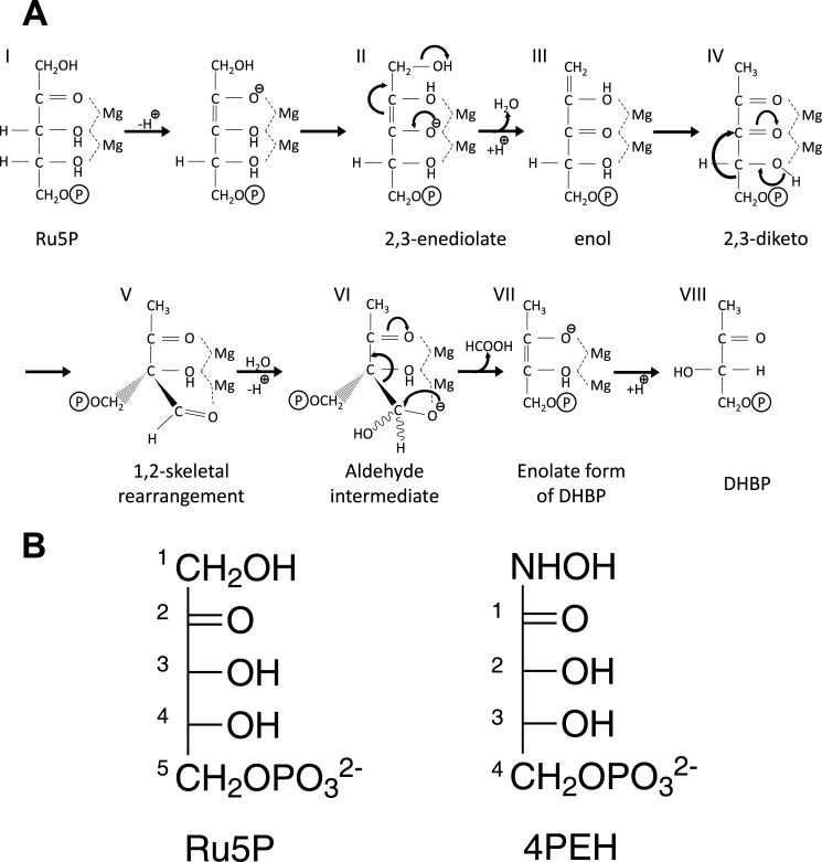
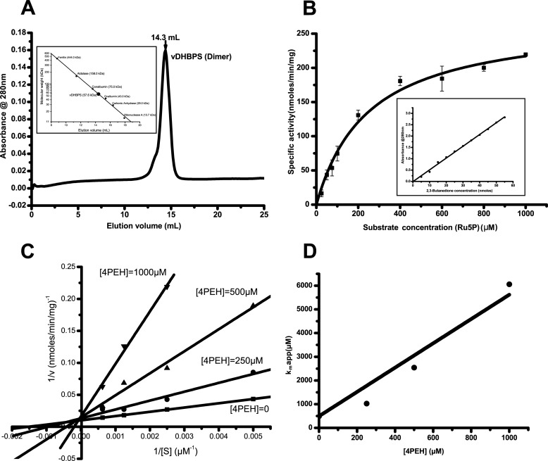
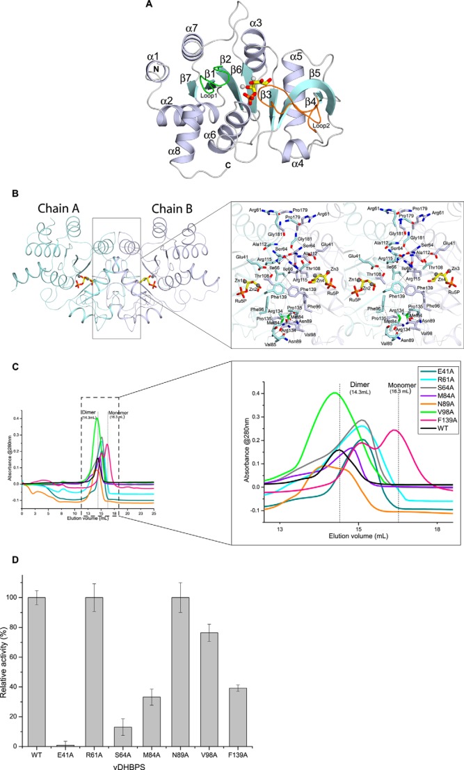
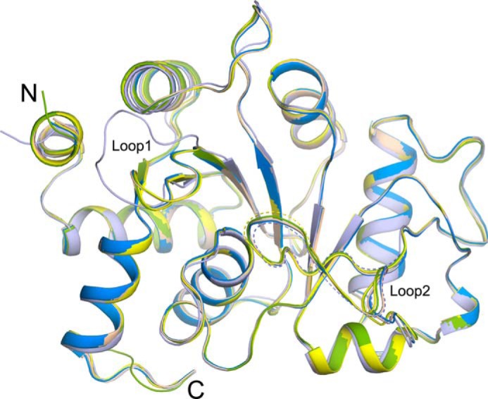
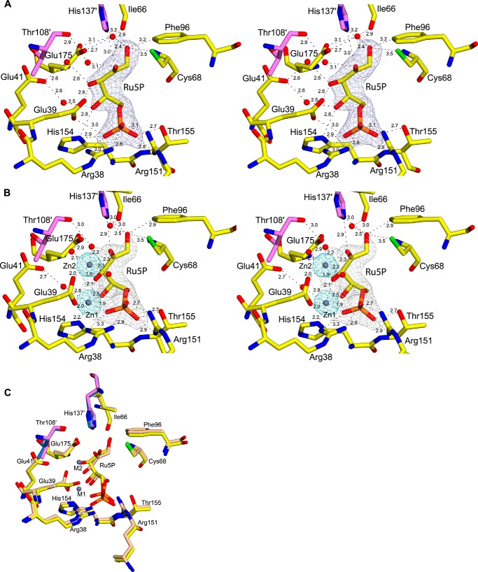
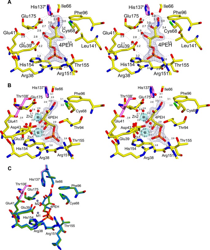
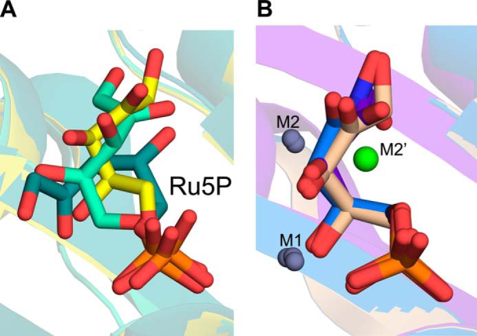
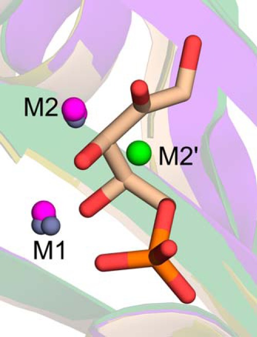
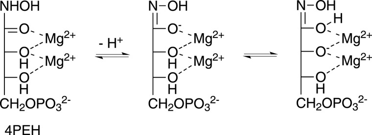
The riboflavin biosynthesis pathway has been shown to be essential in many pathogens and is absent in humans. Therefore, enzymes involved in riboflavin synthesis are considered as potential antibacterial drug targets. The enzyme 3,4-dihydroxy-2-butanone-4-phosphate synthase (DHBPS) catalyzes one of the two committed steps in the riboflavin pathway and converts d-ribulose 5-phosphate (Ru5P) to l-3,4-dihydroxy-2-butanone 4-phosphate and formate. Moreover, DHBPS is shown to be indispensable for Mycobacterium, Salmonella, and Helicobacter species. Despite the essentiality of this enzyme in bacteria, no inhibitor has been identified hitherto. Here, we describe kinetic and crystal structure characterization of DHBPS from Vibrio cholerae (vDHBPS) with a competitive inhibitor 4-phospho-d-erythronohydroxamic acid (4PEH) at 1.86-Å resolution. In addition, we also report the structural characterization of vDHBPS in its apo form and in complex with its substrate and substrate plus metal ions at 1.96-, 1.59-, and 2.04-Å resolution, respectively. Comparison of these crystal structures suggests that 4PEH inhibits the catalytic activity of DHBPS as it is unable to form a proposed intermediate that is crucial for DHBPS activity. Furthermore, vDHBPS structures complexed with substrate and metal ions reveal that, unlike Candida albicans, binding of substrate to vDHBPS induces a conformational change from an open to closed conformation. Interestingly, the position of second metal ion, which is different from that of Methanococcus jannaschii, strongly supports an active role in the catalytic mechanism. Thus, the kinetic and structural characterization of vDHBPS reveals the molecular mechanism of inhibition shown by 4PEH and that it can be explored further for designing novel antibiotics.

Keywords: Crystal Structure; DHBPS; Enzyme Inhibitor; Enzyme Kinetics; Enzyme Mechanism; Riboflavin; Ribulose 5-Phosphate; ribB.


Figures

FIGURE 1.
FIGURE 1.
Reaction catalyzed by DHBPS. A, proposed mechanism for the conversion of Ru5P to DHBP by DHBPS (18). B, chemical structures of substrate Ru5P and inhibitor 4PEH.
FIGURE 2.
FIGURE 2.
Oligomeric and kinetic characterization of vDHBPS. A, size exclusion chromatography elution profile of vDHBPS with a Superdex 200 column. The inset shows the elution profile of standard molecular masses from the same column. B, Michaelis-Menten plot for vDHBPS with various substrate (Ru5P) concentrations. Error bars represent S.D. The inset shows the standard plot generated with known concentration of 2,3-butanedione and used for converting absorbance to DHBP. C, Lineweaver-Burk plot for different concentrations of substrate (Ru5P) at each inhibitor (4PEH) concentration. D, the calculated apparent Km is plotted against inhibitor (4PEH) concentration to estimate Ki.
FIGURE 3.
FIGURE 3.
Structural characterization of vDHBPS. A, the overall structure of vDHBPS. Secondary structures are shown in white-blue for α-helix, cyan for β-strands, and white for loops. Ru5P and metal ions are represented as stick and sphere models, respectively. Loop1 and Loop2 are shown in green and orange, respectively. B, the association of vDHBPS as a dimer. Each monomer is shown in different colors. The Ru5P and metal ions are shown at the corresponding active site of each monomer. The closer view shows the amino acid residues involved in dimerization, and their interactions are shown in dashed lines. C, size exclusion chromatography elution profile of different mutants of vDHBPS from a Superdex 200 column. D, the relative catalytic activity of different mutants of vDHBPS with respect to wild-type (WT) vDHBPS. Error bars represent S.D.
FIGURE 4.
FIGURE 4.
Conformational changes among vDHBPS structures. Superposition of crystal structures of vDHBPS in the apo form (light blue) and in complex with Ru5P (yellow), Ru5P plus zinc ions (wheat), 4PEH (green), and 4PEH plus zinc ions (marine blue) is shown. Dashed lines indicate lack of electron density in that region and not included in the model.
FIGURE 5.
FIGURE 5.
Substrate binding site of vDHBPS. A, stereodiagram showing a 2FoFc electron density map contoured at 1.0 σ for Ru5P. Residues interacting with Ru5P are displayed along with their distances. Residues from the other monomer are shown in magenta. B, stereodiagram showing a 2FoFc electron density map for Ru5P and Zn2+ contoured at 1.5 and 5.0 σ, respectively. The distances are shown for the close residues. C, superposition of the active site of E-Ru5P (yellow) and E-Ru5P-Zn2+ (wheat).
FIGURE 6.
FIGURE 6.
Inhibitor binding site of vDHBPS. A, stereodiagram showing a 2FoFc electron density map contoured at 1.2 σ for 4PEH. Residues interacting with 4PEH are displayed along with their distances, and residues from the other monomer are shown in magenta. B, stereodiagram showing an electron density map for 4PEH and Zn2+ contoured at 2.0 and 8.0 σ, respectively. The distances are shown for the close residues. C, superposition of the active site of E-4PEH (green) and E-4PEH-Zn2+ (marine blue).
FIGURE 7.
FIGURE 7.
Comparison of vDHBPS with other homologs. A, superposition of crystal structures of DHBPS from V. cholerae (wheat), S. typhimurium (olive green), M. tuberculosis (gray), M. jannaschii (purple), M. grisea (forest green), E. coli (brown), S. pneumoniae (orange), and C. albicans (deep teal). B, multiple sequence alignment of vDHBPS. Sequences of DHBPS from known structures are aligned using Clustal Omega (61). The secondary structures are displayed on the top of the alignment for vDHBPS. Identical residues are shown in white with a red background, whereas similar residues are shown in red. The figure was generated through ESPript (62).
FIGURE 8.
FIGURE 8.
Comparison of conformations of Ru5P. A, superposition of Ru5P from V. cholerae (yellow), S. typhimurium (green-cyan), and C. albicans (deep teal) in the absence of metal ions. B, superposition of Ru5P and 4PEH in the presence of metal ions (M1, M2, and M2′) from E-Ru5P-Zn2+ (wheat), E-4PEH-Zn2+ (marine blue), and M. jannaschii (purple-blue).
FIGURE 9.
FIGURE 9.
Comparison of position of metal ions in DHBPS. Superposition of two Zn2+ (M1 and M2; gray) from E-Ru5P-Zn2+, one Zn2+ (M1; gray), and one Mg2+ (M2; purple) from S. typhimurium and two Mg2+ (M1 and M2; purple) from M. grisea, one Zn2+ (M1; gray), and one Ca2+ (M2′; green) from M. jannaschii is shown. The Ru5P from E-Ru5P-Zn2+ is shown in sticks for reference. The M1 and M2 positions are observed in all three species, whereas M2′ is observed only in M. jannaschii.
FIGURE 10.
FIGURE 10.
Proposed mechanism of inhibition by 4PEH. Hydroxamic acids can exist in hydroximic acid form or hydroximate following nitrogen deprotonation. The 4PEH may exist as a stable hydroximate complex with Mg2+ that is unable to induce dehydration of the NOH group and thus leads to inhibition.

References

    1. Bacher A. (1991) in Chemistry and Biochemistry of Flavoenzymes (Müller F., ed) Vol. I, pp. 349–370, CRC Press, Boca Raton, FL
    1. Cole S. T., Eiglmeier K., Parkhill J., James K. D., Thomson N. R., Wheeler P. R., Honoré N., Garnier T., Churcher C., Harris D., Mungall K., Basham D., Brown D., Chillingworth T., Connor R., Davies R. M., Devlin K., Duthoy S., Feltwell T., Fraser A., Hamlin N., Holroyd S., Hornsby T., Jagels K., Lacroix C., Maclean J., Moule S., Murphy L., Oliver K., Quail M. A., Rajandream M. A., Rutherford K. M., Rutter S., Seeger K., Simon S., Simmonds M., Skelton J., Squares R., Squares S., Stevens K., Taylor K., Whitehead S., Woodward J. R., Barrell B. G. (2001) Massive gene decay in the leprosy bacillus. Nature 409, 1007–1011 - PubMed
    1. Gerdes S. Y., Scholle M. D., D'Souza M., Bernal A., Baev M. V., Farrell M., Kurnasov O. V., Daugherty M. D., Mseeh F., Polanuyer B. M., Campbell J. W., Anantha S., Shatalin K. Y., Chowdhury S. A., Fonstein M. Y., Osterman A. L. (2002) From genetic footprinting to antimicrobial drug targets: examples in cofactor biosynthetic pathways. J. Bacteriol. 184, 4555–4572 - PMC - PubMed
    1. Sassetti C. M., Boyd D. H., Rubin E. J. (2003) Genes required for mycobacterial growth defined by high density mutagenesis. Mol. Microbiol. 48, 77–84 - PubMed
    1. Bacher A., Eberhardt S., Richter G. (1996) in Escherichia coli and Salmonella: Cellular and Molecular Biology (Neidhardt F. C., Ingraham J. L., Low K. B., Magasanik B., Schaechter M., Umbarger H. E., eds), Vol. 1, 2nd Ed., pp. 657–664, ASM Press, Washington, D. C.

Publication types

LinkOut - more resources