

GD3- and O-acetylated GD3-gangliosides in the GM2 synthase-deficient mouse brain and their immunohistochemical localization
- PMID: 25792782
- PMCID: PMC4338816
- DOI: 10.2183/pjab.82.189
GD3- and O-acetylated GD3-gangliosides in the GM2 synthase-deficient mouse brain and their immunohistochemical localization
Abstract
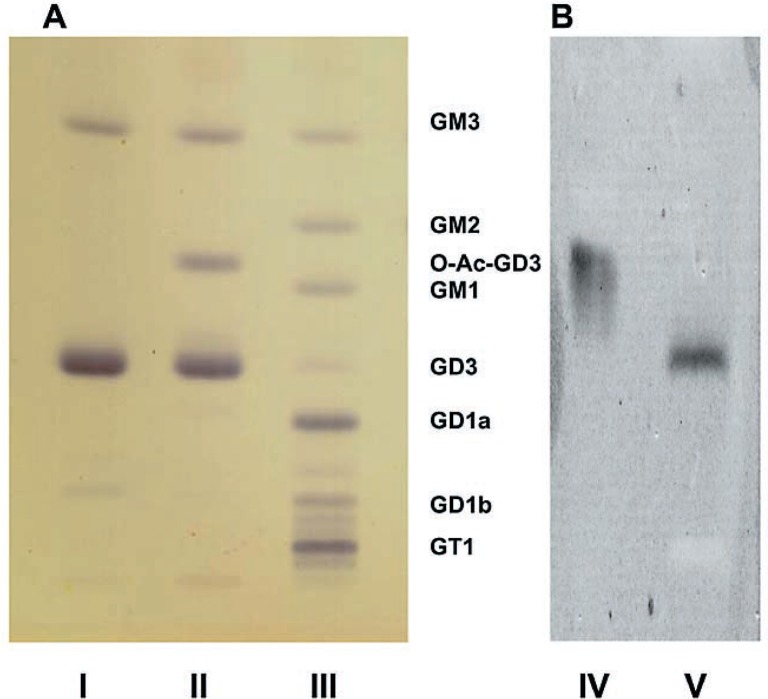
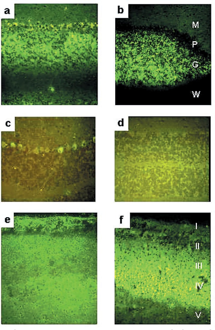
Gangliosides in the brain of the knockout mouse deficient in the activity of β1,4 N-acetylgalactosaminyl transferase (β1,4 GalNAc-T)(GM2 synthase) consisted of nearly exclusively of GM3- and GD3-gangliosides as expected from the known substrate specificity of the enzyme and in confirmation of the initial reports from two laboratories that generated the mutant mouse experimentally. The total molar amount of gangliosides was approximately 30% higher in the mutant mouse brain than that in the wild-type brain. However, contrary to the initial reports, one-fourth of total GD3-ganglioside was O-acetylated. It reacted positively with an anti-O-acetylated GD3 monoclonal antibody and disappeared with a corresponding increase in GD3-ganglioside after mild alkaline treatment. The absence of O-acetylated GD3 in the initial reports can be explained by the saponification step included in their analytical procedures. Although quantitatively much less and identification tentative, we also detected GT3 and O-acetylated GT3. Anti-GD3 and anti-O-acetylated GD3 monoclonal antibodies gave positive reactions in the brain of mutant mouse as expected from the analytical results. Either antibody barely stained wild-type brain except for immunoreactivity of GD3 in the cerebellar Purkinje cells. The distributions of GD3 and O-acetylated GD3 in the brain of mutant mouse were similar but differential localization was noted in the cerebellar Purkinje cells and cerebral cortex.
Keywords: GM2-synthase; O-acetylated ganglioside; Sphingolipid; ganglioside; immunohistochemistry; knockout mouse.
Figures
References
-
- Nagata, Y., Yamashiro, S., Yodoi, J., Lloyd, K. O., Shiku, H., and Furukawa, K. (1992) Expression cloning of β1,4 N-acetylgalactosaminyltransferase cDNAs that determine the expression of GM2 and GD2 gangliosides. J. Biol. Chem. 267, 12082–12089. - PubMed
-
- Sango, K., Johnson, O. N., Kozak, C. A., and Proia, R. L. (1995) β-1,4-N-acetylgalactosaminyltransferase involved in ganglioside synthesis: cDNA sequence, expression, and chromosome mapping of the mouse gene. Genomics 27, 362–365. - PubMed
-
- Takamiya, K., Yamamoto, A., Furukawa, K., Yamashiro, S., Shin, M., Okada, M., Fukumoto, S., and Haraguchi, M., Takeda, N., Fujimura, K.et al. (1996) Mice with disrupted GM2/GD2 synthase gene lack complex gangliosides but exhibit only subtle defects in their nervous system. Proc. Natl. Acad. Sci. USA 93, 10662–10667. - PMC - PubMed