. 2006 Dec;82(9):359-74.
doi: 10.2183/pjab.82.359. Epub 2006 Feb 12.

Molecular systematics and ultrastructural characterization of a forgotten species: Chattonidium setense (Ciliophora, Heterotrichea)

Affiliations

Molecular systematics and ultrastructural characterization of a forgotten species: Chattonidium setense (Ciliophora, Heterotrichea)

Letizia Modeo et al. Proc Jpn Acad Ser B Phys Biol Sci. 2006 Dec.

Abstract

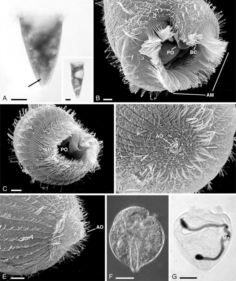
In the present paper we redescribe the ciliate Chattonidium setense Villeneuve 1937 combining morphological observations (live, stained, scanning, and transmission electron microscope) with behavioral notes and molecular data. Ultrastructural analysis revealed remarkable similarities between Chattonidium and representative members of the class Heterotrichea in cortical structure and cytoplasmic organization. The most similar genus for these aspects appears to be Condylostoma. To verify this relatedness, 18S rRNA genes from Chattonidium and from one Condylostoma species were sequenced. Phylogenetic analysis indicates Chattonidium belongs to the class Heterotrichea defined according to the modern taxonomy, and confirms its relatedness with Condylostoma already hypothesized by Villeneuve-Brachon (1940). The presence of the aboral cavity complex, a unique feature never described in other ciliates, and its peculiar organization revealed by ultrastructural analysis fully justify, in our opinion, the maintenance of Chattonidium in the separate family Chattonidiidae, established by Villeneuve-Brachon in 1940.

Keywords: Protozoa; SSU rRNA; heterotrichs; molecular phylogeny; protist; ultrastructure.


Figures

Fig. 1.
Fig. 1.
A–G. General morphology of Chattonidium setense. A. Picture of a live specimen. Arrow indicates the aboral cavity. In the inset a specimen at an early stage of binary fission. Scale bars = 50 μm. B, C. SEM pictures of the apical region. AM, adoral membranelles; BC, bundle of cilia; PO, paraoral membrane; SK, somatic kineties. Scale bars = 10 μm. D, E. The aboral region in a relaxed state (D) and during contraction (E). AO, aboral orifice. Arrows indicate the crowns of longer cilia. Scale bars = 10 μm. F. A contracted specimen in vivo. Scale bar = 50 μm. G. The macronucleus (Ma). Scale bar = 50 μm.
Fig. 2.
Fig. 2.
A–K. Fine structure by TEM. A. The cortical region. Kd, kinetodesmata. Bar = 0.25 μm. B. The different cytoplasmic regions. Ec, ectoplasm; En, endoplasm; FL, fibrous layer; MB, deep microtubular band. Scale bar = 0.25 μm. C–F. Extrusomes. Fs, flask-shaped extrusomes; Mu, mucocyst-like extrusomes. Scale bars = 0.25 μm. G–I. Somatic ciliature. G. Longitudinal section of a dikinetid. Scale bar = 0.25 μm. H, I. Cross-sections. A, anterior kinetosome; LF, lateral fiber; P, posterior kinetosome; Pc, postciliary ribbon; T, transverse ribbon. Arrow indicates the microtubular ribbon connecting the kinetosomes. Scale bars = 0.50 μm. J. Oral ciliature. Pc, postciliary ribbon; T, transverse ribbon. Arrow indicates the microtubular ribbon connecting the kinetosomes. Scale bar = 0.25 μm. K. An oral dikinetid and the bundle of somatic cilia present on the external side of the cytoplasmic expansion covering part of the peristome right margin. AM, adoral membranelles; BC, bundle of cilia. Arrows indicate the nematodesmata. Scale bar = 0.25 μm.
Fig. 3.
Fig. 3.
A–B. Fine structure by TEM. A. Cytoplasmic features. Arrows indicate the numerous paraglycogen granules. Scale bar = 0.5 μm. B. Macronuclear organization. Nu, nucleoli. Scale bar = 1 μm.
Fig. 4.
Fig. 4.
A–D. Fine structure by TEM. The aboral cavity complex. A. Section just below the apical end of the cavity. B. Middle region of the cavity with the six membranelles. C, D. Terminal region of the cavity. Bundles of microtubules originate from the ciliary bases and surround the cavity (Figs. 3A, 4C). The cilia are closer to each other and follow a spiral pattern. BM, bundles of microtubules; F, fibrils; Mu, mucocyst-like extrusomes. Arrow indicates a paraglycogen granule. Scale bars = 2 μm. In the inset of D a semithin, longitudinal section of the cavity. Scale bar = 10 μm.
Fig. 5.
Fig. 5.
Phylogenetic relationships within the class Heterotrichea based on 18S rRNA gene sequences. Phylogenetic analysis performed by DNAPARS program for maximum parsimony (Felsenstein 1989) and limited to 1454 positions. A proper selection of slow evolving 18S rRNA gene sequences belonging to the subphylum Intramacronucleata was chosen as outgroup (dashed box). Numbers at nodes represent the bootstrap values out of 1000 replicates. Lengths of the branches are informative and calculated using the “calculate branch lengths” option in the ARB software package (Ludwig et al. 2004). The scale bar corresponds to 10 substitutions per 100 nucleotide positions. The association of Chattonidium and Condylostoma is highly supported.
Fig. 6.
Fig. 6.
Phylogenetic relationships within the class Heterotrichea based on 18S rRNA gene sequences. Phylogenetic analysis performed by TREEPUZZLE program (Strimmer and von Haeseler 1996), choosing mixed rate heterogeneity (1 invariable plus 8 gamma rates) and the Tamura-Nei model of substitution (Tamura and Nei 1993). Numbers at nodes represent the reliability values. Filter, outgroup, and scale bar like in Fig. 5. A multifurcation close to the base of Heterotrichea is evident.
Fig. 7.
Fig. 7.
The original drawing of Chattonidium setense by Villeneuve-Brachon (1940). Reprinted with permission from CNRS EDITIONS, Archives de Zoologie Expérimentale et Générale, tome 82, 1940, Simone Villeneuve-Brachon, Recherches sur les ciliés hétérotriches: cinétome, argyrome, myonèmes, formes nouvelles ou peu connues, page 75. Scale bar = 50 μm.

References

    1. Bütschli, O. (1889) InKlassen und Ordnungen des Thier-Reichs, wissenschaftlich dargestellt in Wort und Bild (ed. Bronn H. G.). Leipzig, Winter, pp. 1585–2035.
    1. Kahl, A. (1932) InDie Tierwelt Deutschlands und der angrenzenden Meeresteile (ed. Dahl F.). Gustav Fischer, Jena, 25, pp. 399–650.
    1. Corliss, J. O. (1979) The ciliated protozoa. Characterization, classification and guide to the literature 2nd edition Pergamon Press, Oxford-New York-Toronto-Sydney-Paris-Frankfurt.
    1. Gerassimova, Z. P., and Seravin, L. N. (1976) Zool. Zh. 55, 645–656.
    1. Lynn, D. H. (1976a) J. Protozool. 23, 302–314.