

Patients with diabetic retinopathy have high retinal venous pressure
- PMID: 25793018
- PMCID: PMC4365968
- DOI: 10.1186/s13167-015-0027-1
Patients with diabetic retinopathy have high retinal venous pressure
Abstract
Background: The introduction of ophthalmodynamometric measurement of retinal venous pressure (RVP) now permits the quantification, or at least an approximation, of the real pressure in the retinal veins.
Methods: We measured the RVP of healthy control subjects, patients with diabetes without diabetic retinopathy (nonDR) and patients with diabetes and diabetic retinopathy (DR).
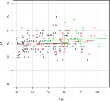
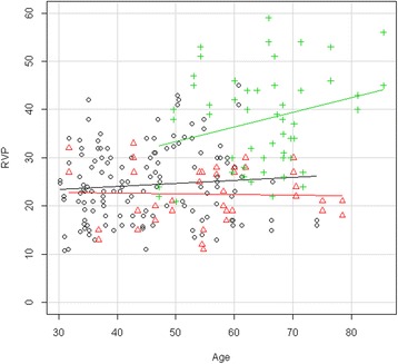
Results: The mean ± SD RVP for the control, nonDR and DR groups were 23.4 ± 7.33, 22.5 ± 5.78 and 37.7 ± 10.1 mmHg, respectively. In the diabetes patients with DR, the RVP was markedly and significantly increased, and this result was significantly age dependent. RVP was not increased in the group of diabetes patients without DR. In our tested population, diabetes had a minor influence on intraocular pressure.
Conclusion: Regardless of the cause, a marked increase in RVP in diabetes patients with DR is clinically relevant, as it reduces perfusion pressure and increases transmural pressure. The reduced perfusion pressure contributes to hypoxia, and the increased transmural pressure can facilitate retinal edema. Diabetes is an increasing burden, and DR is one of its most severe complications. Strategies to recognize the risk for DR and to develop personalized prevention and therapy therefore have major implications.
Trial registration: ClinicalTrials.gov ID: NCT01771835.
Keywords: Contact lens dynamometer (CLD); Diabetic retinopathy (DR); Ocular dynamic force (ODF); Ophthalmodynamometry; Personalized prevention; Retinal venous pressure (RVP); Spontaneous venous pulsation.
Figures


Similar articles
-
Retinal venous pressure: the role of endothelin.EPMA J. 2015 Oct 26;6:21. doi: 10.1186/s13167-015-0043-1. eCollection 2015. EPMA J. 2015. PMID: 26504500 Free PMC article. Review.
-
Retinal venous pressure in the non-affected eye of patients with retinal vein occlusions.Graefes Arch Clin Exp Ophthalmol. 2014 Oct;252(10):1569-71. doi: 10.1007/s00417-014-2617-3. Epub 2014 Mar 28. Graefes Arch Clin Exp Ophthalmol. 2014. PMID: 24676960
-
Retinal venous pressure measurements in patients with Flammer syndrome and metabolic syndrome.EPMA J. 2017 Sep 5;8(4):339-344. doi: 10.1007/s13167-017-0105-7. eCollection 2017 Dec. EPMA J. 2017. PMID: 29209437 Free PMC article.
-
The effect of flammer-syndrome on retinal venous pressure.BMC Ophthalmol. 2014 Oct 13;14:121. doi: 10.1186/1471-2415-14-121. BMC Ophthalmol. 2014. PMID: 25312339 Free PMC article.
-
Perspectives on diabetic retinopathy.Am J Ophthalmol. 2003 Jul;136(1):122-35. doi: 10.1016/s0002-9394(03)00219-8. Am J Ophthalmol. 2003. PMID: 12834680 Review.
Cited by
-
Retinal venous pressure: the role of endothelin.EPMA J. 2015 Oct 26;6:21. doi: 10.1186/s13167-015-0043-1. eCollection 2015. EPMA J. 2015. PMID: 26504500 Free PMC article. Review.
-
The retinal venous pressure at different levels of airway pressure measured with a new method.Graefes Arch Clin Exp Ophthalmol. 2024 Sep;262(9):2971-2976. doi: 10.1007/s00417-024-06483-0. Epub 2024 Apr 9. Graefes Arch Clin Exp Ophthalmol. 2024. PMID: 38592501 Free PMC article.
-
The discovery of the Flammer syndrome: a historical and personal perspective.EPMA J. 2017 May 22;8(2):75-97. doi: 10.1007/s13167-017-0090-x. eCollection 2017 Jun. EPMA J. 2017. PMID: 28725290 Free PMC article. Review.
-
Quantitative analysis of early retinal vascular changes in type 2 diabetic patients without clinical retinopathy by optical coherence tomography angiography.Int Ophthalmol. 2022 Feb;42(2):367-375. doi: 10.1007/s10792-022-02230-8. Epub 2022 Jan 31. Int Ophthalmol. 2022. PMID: 35099665
-
Retinal venous pressure is decreased after anti-VEGF therapy in patients with retinal vein occlusion-related macular edema.Graefes Arch Clin Exp Ophthalmol. 2021 Jul;259(7):1853-1858. doi: 10.1007/s00417-020-05068-x. Epub 2021 Jan 15. Graefes Arch Clin Exp Ophthalmol. 2021. PMID: 33447857 Free PMC article.
References
Associated data
LinkOut - more resources
Full Text Sources
Other Literature Sources
Medical
