Case Reports
. 2015 Jan 18;12(1):e12597.
doi: 10.5812/iranjradiol.12597. eCollection 2015 Jan.

Clinico-radiologic findings in primary cutaneous extranodal natural killer/t-cell lymphoma, nasal type mimicking cellulitis of the left arm

Affiliations
Case Reports

Clinico-radiologic findings in primary cutaneous extranodal natural killer/t-cell lymphoma, nasal type mimicking cellulitis of the left arm

Soo Hyun Kim et al. Iran J Radiol. .

Abstract

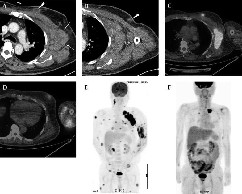
Extranodal natural killer (NK)/T-cell lymphoma is a very rare and aggressive disease characterized histopathologically by an Epstein-Barr virus (EBV)-positive atypical lymphoid cytotoxic infiltrate, extensive vascular destruction, and prominent tissue necrosis. It commonly shows cutaneous lesions that primarily or secondarily mimic cellulitis at the primary site. We report on a very rare case of extranodal NK/T-cell lymphoma, nasal type of skin/soft tissue, in a 64-year-old man, and describe the radiological findings. The condition was misdiagnosed as cellulitis of the left arm based on initial noninvasive clinical and radiologic work-up.

Keywords: Cellulitis; Lymphoma; Natural Killer T-Cell.


Figures

Figure 1.
Figure 1.. A 64-year-old man with a 3-month history of painful swelling and redness of the left upper arm. Cutaneous manifestation of primary cutaneous extranodal NK/T-cell lymphoma, nasal type. (A, B,) There was erythematous to violaceous swelling, nodules, and ulceration with a crust on the left arm.
Figure 2.
Figure 2.. Magnetic resonance imaging and contrast-enhanced computed tomography findings of the left upper arm in a case of primary cutaneous extranodal NK/T-cell lymphoma, nasal type. Fat-suppressed T2-weighted (A) and contrast-enhanced fat suppressed T1-weighted (B) MRI images showed diffuse soft tissue infiltration and subcutaneous edema, with intermediate to subtle low-signal-intensity areas in the T2-weighted image (arrows in A) and heterogeneous enhancement (arrows in B) in the medial aspect of the left upper arm. Enhanced CT images (C, D, and E) of the left humerus (C-E) also showed diffuse soft tissue infiltration (arrows in C) and mixed subcutaneous nodules (arrowheads in D and E) with poorly enhancing peripheral rims and relatively radiolucent central areas.
Figure 3.
Figure 3.. Left axillary lymphadenopathies and cutaneous involvement in a case of primary cutaneous extranodal NK/T-cell lymphoma, nasal type. A and B, Axial contrast-enhanced CT images showed small discrete subcutaneous nodules with poorly enhanced peripheral rims and relatively radiolucent central areas (arrowheads) in the left anterior chest wall, and multiple conglomerated lymphadenopathies in the left axilla (arrows). C and D, These nodules and axillary lymphadenopathies showed high fludeoxyglucose (FDG) uptake in PET-CT images. E, Coronal maximum-intensity-projection PET image showing multiple 18 F-FDG-avid lesions. F, Follow-up PET image after seven months showing regression of multiple 18 F-FDG-avid lesions. The remaining high-FDG-uptake lesion in the left upper lung (arrow) was caused by pneumonic infiltration.
Figure 4.
Figure 4.. Pathological specimens. A, Atypical lymphoid cell infiltration in the dermis and subcutaneous deep fat layer, with extensive necrosis (H and E staining, magnification × 10). B, Angiodestruction by tumor cells (H and E staining, magnification × 200). C, Immunohistochemical staining showing that lymphoma cells were positive for CD56. D, Granzyme B. E, Epstein-Barr virus in situ.

Similar articles

Cited by

References

    1. Tomonaga M. [Outline and direction of revised WHO classification of Tumors of Haematopoietic and Lymphoid Tissues]. Rinsho Ketsueki. 2009;50(10):1401–6. - PubMed
    1. Choi YL, Park JH, Namkung JH, Lee JH, Yang JM, Lee ES, et al. Extranodal NK/T-cell lymphoma with cutaneous involvement: 'nasal' vs. 'nasal-type' subgroups--a retrospective study of 18 patients. Br J Dermatol. 2009;160(2):333–7. doi: 10.1111/j.1365-2133.2008.08922.x. - DOI - PubMed
    1. Pagano L, Gallamini A, Trape G, Fianchi L, Mattei D, Todeschini G, et al. NK/T-cell lymphomas 'nasal type': an Italian multicentric retrospective survey. Ann Oncol. 2006;17(5):794–800. doi: 10.1093/annonc/mdl015. - DOI - PubMed
    1. Stokkermans-Dubois J, Jouary T, Vergier B, Delaunay MM, Taieb A. A case of primary cutaneous nasal type NK/T-cell lymphoma and review of the literature. Dermatology. 2006;213(4):345–9. doi: 10.1159/000096201. - DOI - PubMed
    1. Berti E, Recalcati S, Girgenti V, Fanoni D, Venegoni L, Vezzoli P. Cutaneous extranodal NK/T-cell lymphoma: a clinicopathologic study of 5 patients with array-based comparative genomic hybridization. Blood. 2010;116(2):165–70. doi: 10.1182/blood-2009-11-252957. - DOI - PubMed

Publication types

LinkOut - more resources