

Multimodality imaging of left circumflex artery to coronary sinus fistula
- PMID: 25793089
- PMCID: PMC4349104
- DOI: 10.5812/iranjradiol.6878
Multimodality imaging of left circumflex artery to coronary sinus fistula
Abstract
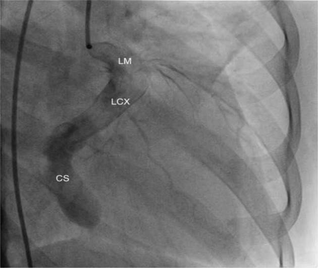
Coronary artery fistula (CAF) is a rare anomaly of the coronary artery. Patients with this condition are usually asymptomatic. However, cardiac failure may occur later in life due to progressive enlargement of the fistula. Diagnosis is traditionally made by echocardiogram and conventional angiogram. However with the advantage of new technologies such as computed tomography (CT) coronary angiography, the course and communications of these fistulae can be delineated non-invasively and with greater accuracy. We report a case of a left circumflex artery fistula to the coronary sinus which was suspected on echocardiogram and the diagnosis was clinched on ECG-gated CT.
Keywords: Circumflex Artery; Computed tomography; Coronary Sinus; Fistula.
Figures


References
-
- Krause W. [Uber den ursprung einer accessorischen a Coronaria aus der a.] Z Ratl Med Pulmonalis. 1865;24:225–7.
-
- Wenger NK. In: Rare causes of coronary artery disease. JW Hurst, editor. New York: McGraw-Hill; 1978.
-
- Lin FC, Chang HJ, Wen MS, Yeh SJ, Wu D. Multiplane transesophageal echocardiography in the diagnosis of congenital coronary artery fistula. Am Heart J. 1995;130(6):1236–42. - PubMed
-
- Hirooka K, Hanatani A, Nakatani S, Yasumura Y, Bando K, Miyatake K, et al. Huge saccular aneurysm in a coronary-pulmonary fistula fed by the left and right coronary arteries. Circ J. 2002;66(5):525–7. - PubMed
Publication types
LinkOut - more resources
Full Text Sources
Other Literature Sources