

Atlanto-occipital dislocation
- PMID: 25793163
- PMCID: PMC4363805
- DOI: 10.5312/wjo.v6.i2.236
Atlanto-occipital dislocation
Abstract
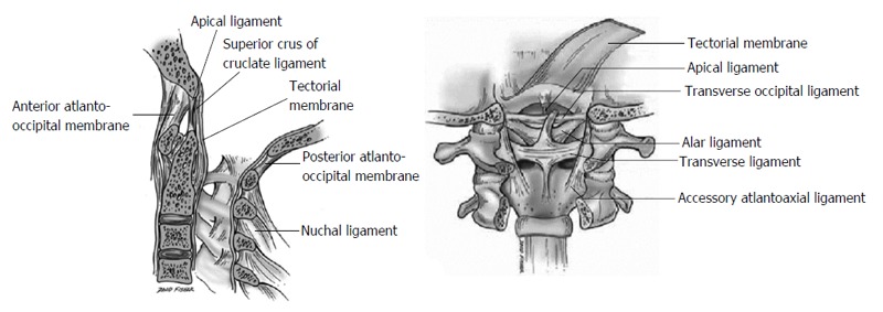
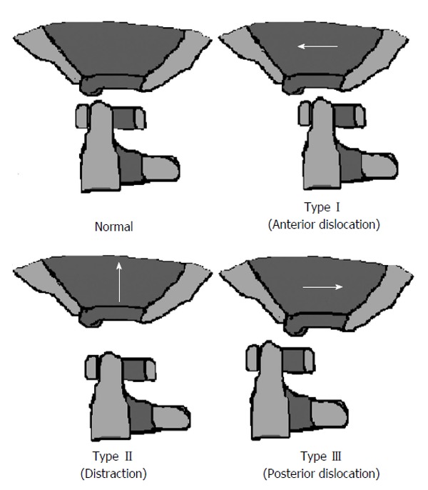
Atlanto-occipital dislocation (AOD) is being increasingly recognized as a potentially survivable injury as a result of improved prehospital management of polytrauma patients and increased awareness of this entity, leading to earlier diagnosis and more aggressive treatment. However, despite overall improved outcomes, AOD is still associated with significant morbidity and mortality. The purpose of this paper is to review the biomechanical aspects, clinical features, radiologic criteria, and treatment strategies of AOD. Given that the diagnosis of AOD can be very challenging, a high degree of clinical suspicion is essential to ensure timely recognition and treatment, thus preventing neurological decline or death.
Keywords: Atlanto-occipital dislocation; Cervical spine; Craniocervical junction; Occipitocervical fusion; Trauma.
Figures
References
-
- Bohlman HH. Acute fractures and dislocations of the cervical spine. An analysis of three hundred hospitalized patients and review of the literature. J Bone Joint Surg Am. 1979;61:1119–1142. - PubMed
-
- Payer M, Sottas CC. Traumatic atlanto-occipital dislocation: presentation of a new posterior occipitoatlantoaxial fixation technique in an adult survivor: technical case report. Neurosurgery. 2005;56:E203; discussion E203. - PubMed
-
- Jeszenszky D, Fekete TF, Lattig F, Bognár L. Intraarticular atlantooccipital fusion for the treatment of traumatic occipitocervical dislocation in a child: a new technique for selective stabilization with nine years follow-up. Spine (Phila Pa 1976) 2010;35:E421–E426. - PubMed
-
- Blackwood NJ. III. Atlo-Occipital Dislocation: A Case of Fracture of the Atlas and Axis, and Forward Dislocation of the Occiput on the Spinal Column, Life being Maintained for Thirty-four Hours and Forty Minutes by Artificial Respiration, during which a Laminectomy was Performed upon the Third Cervical Vertebra. Ann Surg. 1908;47:654–658. - PMC - PubMed
-
- Alker GJ, Oh YS, Leslie EV, Lehotay J, Panaro VA, Eschner EG. Postmortem radiology of head neck injuries in fatal traffic accidents. Radiology. 1975;114:611–617. - PubMed
Publication types
LinkOut - more resources
Full Text Sources
Other Literature Sources
