Unusually large erupted complex odontoma: A rare case report
- PMID: 25793183
- PMCID: PMC4362991
- DOI: 10.5624/isd.2015.45.1.49
Unusually large erupted complex odontoma: A rare case report
Abstract
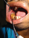
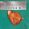
Odontomas are nonaggressive, hamartomatous developmental malformations composed of mature tooth substances and may be compound or complex depending on the extent of morphodifferentiation or on their resemblance to normal teeth. Among them, complex odontomas are relatively rare tumors. They are usually asymptomatic in nature. Occasionally, these tumors become large, causing bone expansion followed by facial asymmetry. Odontoma eruptions are uncommon, and thus far, very few cases of erupted complex odontomas have been reported in the literature. Here, we report the case of an unusually large, painless, complex odontoma located in the right posterior mandible.
Keywords: Facial Asymmetry; Mandible; Odontogenic Tumors; Odontoma.
Figures


References
-
- Reichart P, Philipsen HP. Odontogenic tumours and allied lesions. London: Quintessence Pub; 2004. pp. 141–147.
-
- Vengal M, Arora H, Ghosh S, Pai KM. Large erupting complex odontoma: a case report. J Can Dent Assoc. 2007;73:169–173. - PubMed
-
- Dua N, Kapila R, Trivedi A, Mahajan S, Gupta SD. An unusual case of erupted composite complex odontoma. J Dent Sci Res. 2011;2:1–5.
-
- Serra-Serra G, Berini-Aytes L, Gay-Escoda C. Erupted odontomas: a report of three cases and review of literature. Med Oral Patol Oral Cir Bucal. 2009;14:E299–E303. - PubMed
-
- Garcia-Consuegra L, Junquera LM, Albertos JM, Rodriguez Odontomas. A clinical-histological and retrospective epidemiological study of 46 cases. Med Oral. 2000;5:367–372. - PubMed
Publication types
LinkOut - more resources
Full Text Sources
Other Literature Sources

