. 2015 Mar 20;10(3):e0121534.
doi: 10.1371/journal.pone.0121534. eCollection 2015.

Microvesicles derived from human umbilical cord mesenchymal stem cells facilitate tubular epithelial cell dedifferentiation and growth via hepatocyte growth factor induction

Affiliations

Microvesicles derived from human umbilical cord mesenchymal stem cells facilitate tubular epithelial cell dedifferentiation and growth via hepatocyte growth factor induction

Guan-qun Ju et al. PLoS One. .

Abstract

During acute kidney injury (AKI), tubular cell dedifferentiation initiates cell regeneration; hepatocyte growth factor (HGF) is involved in modulating cell dedifferentiation. Mesenchymal stem cell (MSC)-derived microvesicles (MVs) deliver RNA into injured tubular cells and alter their gene expression, thus regenerating these cells. We boldly speculated that MVs might induce HGF synthesis via RNA transfer, thereby facilitating tubular cell dedifferentiation and regeneration. In a rat model of unilateral AKI, the administration of MVs promoted kidney recovery. One of the mechanisms of action is the acceleration of tubular cell dedifferentiation and growth. Both in vivo and in vitro, rat HGF expression in damaged rat tubular cells was greatly enhanced by MV treatment. In addition, human HGF mRNA present in MVs was delivered into rat tubular cells and translated into the HGF protein as another mechanism of HGF induction. RNase treatment abrogated all MV effects. In the in vitro experimental setting, the conditioned medium of MV-treated injured tubular cells, which contains a higher concentration of HGF, strongly stimulated cell dedifferentiation and growth, as well as Erk1/2 signaling activation. Intriguingly, these effects were completely abrogated by either c-Met inhibitor or MEK inhibitor, suggesting that HGF induction is a crucial contributor to the acceleration of cell dedifferentiation and growth. All these findings indicate that MV-induced HGF synthesis in damaged tubular cells via RNA transfer facilitates cell dedifferentiation and growth, which are important regenerative mechanisms.


Conflict of interest statement

Competing Interests: The authors have declared that no competing interests exist.

Figures

Fig 1
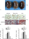
Fig 1. MV administration reverses the abnormal kidney structure and function elicited by AKI at 2 wk post-injury.
(A) Representative micrographs of injured kidneys. In AKI animals treated with RNase-MVs or vehicle, the damaged kidneys display a mottled color, in contrast to a uniform color on the surfaces of the kidneys from MV-treated animals. (B) Representative micrographs illustrating α-SMA staining and Masson’s tri-chrome staining. Weaker positive staining was observed for α-SMA and for collagen on kidney sections from AKI animals receiving MV treatment compared with animals treated with RNase-MVs or vehicle. Magnification, ×40. (C) Serum creatinine value at 2 wk post-injury. Ischemic injury led to a significant increase in the serum creatinine level at 2 wk post-injury, which was inhibited by MV treatment. All quantitative data were obtained from 6 different animals for each experimental condition. *P<0.01, AKI+MVs vs. AKI+VEHICLE; #P<0.01, AKI+RNase-MVs vs. AKI+MVs; xP<0.001, AKI+VEHICLE vs. SHAM. (D) BUN value at 2 wk post-injury. MV administration also greatly inhibited the increase in the BUN level, which occurred at 2 wk post-injury. *P<0.05, AKI+MVs vs. AKI+VEHICLE; #P<0.05, AKI+RNase-MVs vs. AKI+MVs; xP<0.001, AKI+VEHICLE vs. SHAM.
Fig 2
Fig 2. MV administration promotes tubular cell dedifferentiation and proliferation at 48 h post-injury, whereas cell apoptosis is inhibited.
Representative micrographs showing vimentin, PCNA and TUNEL staining of tubular cells. Immuno-staining for vimentin and PCNA proteins, which are indictors for tubular cell dedifferentiation and cell proliferation, respectively, was employed. TUNEL staining was used to detect cell apoptosis. In contrast to the rats treated with vehicle or with RNase-MVs, the rats receiving MV treatment displayed more PCNA- and vimentin-positive stained tubular cells and fewer TUNEL-positive cells on kidney tissue sections. Magnification, ×40.
Fig 3
Fig 3. At 48 h post-injury, kidney HGF gene and protein expression is substantially enhanced by MV administration.
(A)-(D) HGF gene expression in injured kidney tissues. MV administration led to a significant up-regulation of kidney HGF gene expression. The examination of rat HGF expression in kidney tissues using species-specific primers also indicated a similar result. As negative controls, no rat HGF mRNA was identified in MVs or in the cells of origin (hUC-MSCs). RNase pretreatment abolished the effect of MVs. By contrast, EGF, IGF-1 or TGFβ1 gene expression was not altered by MV administration. Gene expression levels in sham-treated samples were regarded as the baseline levels (dotted line). The relative expression levels of each gene were calculated using the 2−ΔΔCt method. The data were collected from 6 rats for each experimental condition. *P<0.05, AKI+MVs vs. AKI+VEHICLE; #P<0.05, AKI+RNase-MVs vs. AKI+MVs. (E) Densitometric analysis of kidney HGF protein expression. At 48 h, MV administration also resulted in a prominent increase in kidney HGF protein expression. This effect was abrogated by RNase pre-treatment. The values in the graph are expressed as densitometric ratios of HGF/GAPDH as fold changes compared with the control (sham-operated samples) (dotted line). *P<0.05, AKI+MVs vs. AKI+VEHICLE; #P<0.05, AKI+RNase-MVs vs. AKI+MVs; xP<0.01, AKI+VEHICLE vs. SHAM. (F) Representative gel photograph of kidney HGF protein expression. (G) HGF staining on kidney sections. Most of the positive staining was observed in damaged tubular cells. HGF staining of injured tubular cells was remarkably intensified in MV-treated animals at 48 h post-injury. Magnification, ×40.
Fig 4
Fig 4. In the scenario of hypoxia/re-oxygenation, rat HGF expression in cultured tubular cells is significantly induced by MV administration.
(A)-(E) HGF gene expression in damaged rat tubular cells. HGF gene expression in injured rat tubular cells was significantly enhanced by MV addition. The examination of rat HGF gene expression using species-specific primers (rat) also revealed a similar result. By contrast, EGF, IGF-1 or TGF-β1 gene expression remained unchanged by MV administration. The gene expression levels in the vehicle-treated cell samples were regarded as the baseline levels (dotted line). The relative expression levels of each gene were calculated using the 2−ΔΔCt method. The data were collected from 5 independent experiments. *P<0.05, MVs vs. VEHICLE; #P<0.05, RNase-MVs vs. MVs. (F) Rat HGF level in the conditioned medium (CM) of tubular cells. Compared with vehicle or with RNase-MVs, MVs caused a remarkable increase in the rat HGF level in the CM after 24 or 48 h of incubation with tubular cells. TECs: tubular epithelial cells. *P<0.02, MVs vs. VEHICLE; #P<0.05, RNase-MVs vs. MVs. (G) Densitometric analysis of HGF protein expression. At 48 h of incubation with tubular cells, MVs resulted in a marked increase in HGF protein expression. The values in the graph are expressed as densitometric ratios of HGF/β-actin as fold changes compared with the control (vehicle-treated cell samples) (dotted line). *P<0.01, MVs vs. VEHICLE; #P<0.01, RNase-MVs vs. MVs. (H) Representative gel photograph of HGF protein expression. (I) HGF staining in injured tubular cells. Stronger HGF-positive staining was observed in tubular cells incubated with MVs compared with RNase-MVs or vehicle, particularly after 48 h of incubation.
Fig 5
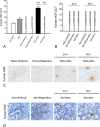
Fig 5. Either in vivo or in vitro, human HGF mRNA present in MVs enters injured rat tubular cells and is translated into the corresponding protein.
(A) Human HGF gene expression in cultured rat tubular cells. The human HGF gene transcript was detectable in MVs and in the cells of origin. After 24 h of incubation with MVs, the human HGF mRNA was present in injured rat tubular cells, whereas human HGF mRNA was absent in cells incubated with vehicle or with RNase-MVs. The Ct for human HGF and β-actin (rat or human) was determined for each sample. The data were collected from 5 independent experiments. TECs: tubular epithelial cells. (B) Human HGF gene expression in rat kidney tissues. The human HGF mRNA was not detected in the affected kidney tissues of MV-treated AKI animals at any given points in time. The Ct for human HGF and rat β-actin was determined for each kidney sample. The data were collected from 3 samples for each group. (C) Human HGF in vitro staining. After MV exposure for 24 or 48 h, a few injured rat tubular cells displayed human HGF-positive expression in the cytoplasm. As a control, no positive staining was observed in cells exposed to vehicle or RNase-MVs. Magnification, ×20. (D) Human HGF in vivo staining. At 24 or 48 h following MV administration, the human HGF protein was detected in a few tubular cells. No positive-stained kidney cells were identifiable in animals treated with vehicle or with RNase-MVs. Magnification, ×40. AKI: acute kidney injury.
Fig 6
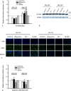
Fig 6. The CM of MV-treated injured tubular cells promotes cell proliferation and inhibits apoptosis.
(A) Densitometric analysis of PCNA protein in tubular cells. The CM of injured tubular cells treated with MVs for 48 h elicited a marked increase in PCNA protein expression in tubular cells. The values in the graph are expressed as densitometric ratios of PCNA/β-actin as fold changes compared with the control (BLANK) (dotted line). CM: conditioned medium of tubular cells; BLANK: medium without exposure to injured tubular cells. *P<0.01, MVs vs. VEHICLE; #P<0.05, RNase-MVs vs. MVs; xP<0.05, VEHICLE vs. BLANK. (B) Representative gel photograph of PCNA protein expression. (C) TUNEL staining. In the presence of the CM of MV-treated tubular cells, few TUNEL-positive cells were observed under ×20 magnification. (D) Quantitative evaluation of cell apoptosis. Cell apoptosis was substantially inhibited by the CM of MV-treated tubular cells. The values in the graph are expressed as ratios of apoptotic cells/total cells as fold changes compared with the control (BLANK) (dotted line). *P<0.01, MVs vs. VEHICLE; #P<0.05, RNase-MVs vs. MVs.
Fig 7
Fig 7. c-Met inhibitor or MEK inhibitor addition abrogates tubular cell dedifferentiation and growth, as well as Erk1/2 signaling activation induced by the CM of MV-treated injured tubular cells.
(A), (C) Densitometric analysis of vimentin, PCNA and p-Erk1/2 protein levels in tubular cells. c-Met inhibitor or MEK inhibitor addition completely abrogated the up-regulation of vimentin, PCNA and p-Erk1/2 protein expression induced by CM of damaged tubular cells treated with MVs. Values in the graph are expressed as densitometric ratios of vimentin/β-actin, PCNA/β-actin or p-Erk1/2/β-actin as fold changes compared with the control (CM of tubular cells treated with vehicle) (dotted line). *P<0.05, MVs vs. VEHICLE; #P<0.05, MVs+PF2341066 or U0126 vs. MVs; xP<0.05, VEHICLE+PF2341066 or U0126 vs. VEHICLE. (B), (D) Representative gel photograph of vimentin, PCNA and p-Erk1/2 protein expression.

References

    1. Ishani A, Xue JL, Himmelfarb J, Eggers PW, Kimmel PL, Molitoris BA, et al. Acute kidney injury increases risk of ESRD among elderly. Journal of the American Society of Nephrology: JASN. 2009;20(1):223–8. 10.1681/ASN.2007080837 - DOI - PMC - PubMed
    1. Venkatachalam MA, Griffin KA, Lan R, Geng H, Saikumar P, Bidani AK. Acute kidney injury: a springboard for progression in chronic kidney disease. American journal of physiology Renal physiology. 2010;298(5):F1078–94. 10.1152/ajprenal.00017.2010 - DOI - PMC - PubMed
    1. Chawla LS, Amdur RL, Amodeo S, Kimmel PL, Palant CE. The severity of acute kidney injury predicts progression to chronic kidney disease. Kidney international. 2011;79(12):1361–9. 10.1038/ki.2011.42 - DOI - PMC - PubMed
    1. Macedo E, Bouchard J, Mehta RL. Renal recovery following acute kidney injury. Current Opinion in Critical Care. 2008;14(6):660–5. 10.1097/MCC.0b013e328317ee6e - DOI - PubMed
    1. Bonventre JV. Pathophysiology of AKI: injury and normal and abnormal repair. Contributions to nephrology. 2010;165:9–17. 10.1159/000313738 - DOI - PubMed

Publication types

MeSH terms