. 2015 Mar 20;11(3):e1005083.
doi: 10.1371/journal.pgen.1005083. eCollection 2015 Mar.

Neurospora importin α is required for normal heterochromatic formation and DNA methylation

Affiliations

Neurospora importin α is required for normal heterochromatic formation and DNA methylation

Andrew D Klocko et al. PLoS Genet. .

Abstract

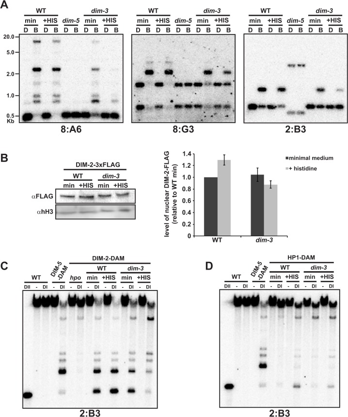
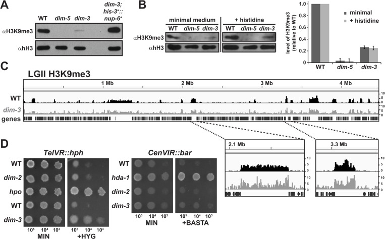
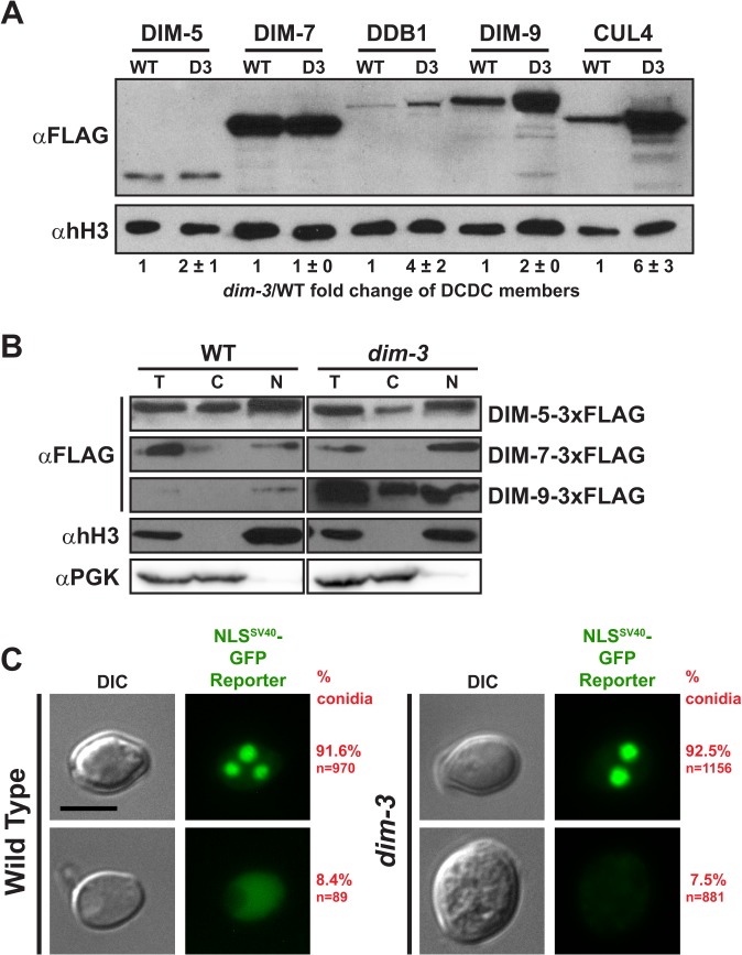
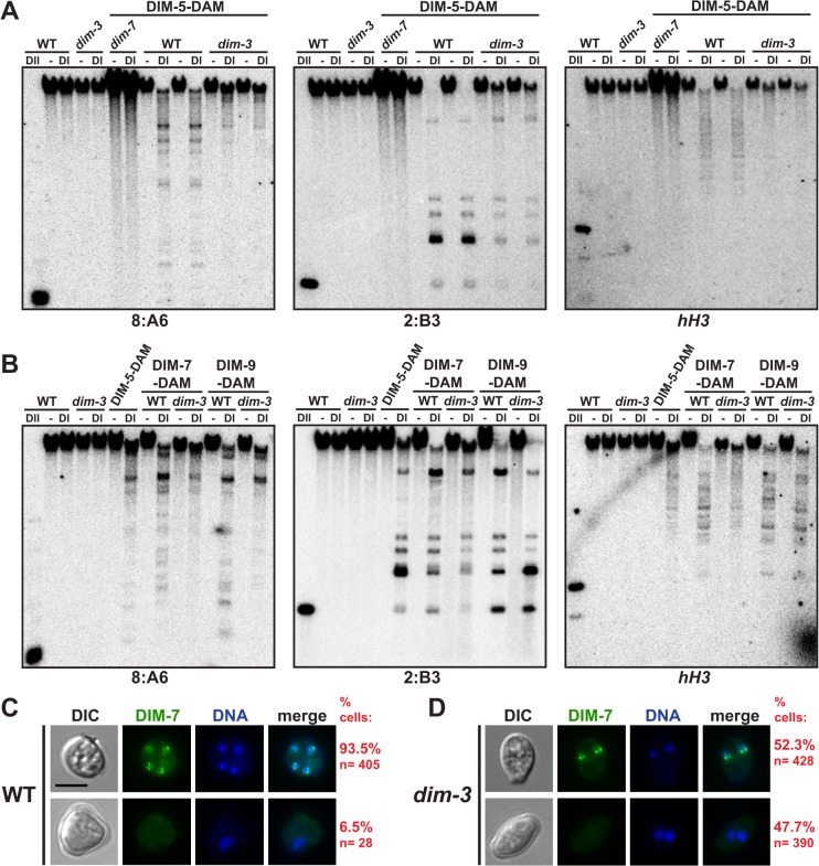
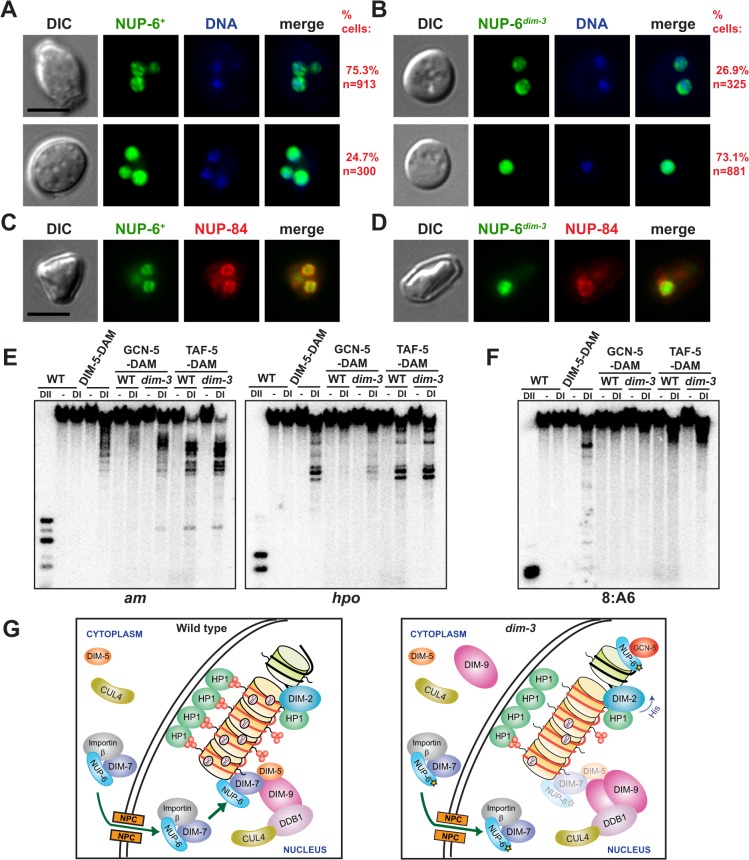
Heterochromatin and associated gene silencing processes play roles in development, genome defense, and chromosome function. In many species, constitutive heterochromatin is decorated with histone H3 tri-methylated at lysine 9 (H3K9me3) and cytosine methylation. In Neurospora crassa, a five-protein complex, DCDC, catalyzes H3K9 methylation, which then directs DNA methylation. Here, we identify and characterize a gene important for DCDC function, dim-3 (defective in methylation-3), which encodes the nuclear import chaperone NUP-6 (Importin α). The critical mutation in dim-3 results in a substitution in an ARM repeat of NUP-6 and causes a substantial loss of H3K9me3 and DNA methylation. Surprisingly, nuclear transport of all known proteins involved in histone and DNA methylation, as well as a canonical transport substrate, appear normal in dim-3 strains. Interactions between DCDC members also appear normal, but the nup-6(dim-3) allele causes the DCDC members DIM-5 and DIM-7 to mislocalize from heterochromatin and NUP-6dim-3 itself is mislocalized from the nuclear envelope, at least in conidia. GCN-5, a member of the SAGA histone acetyltransferase complex, also shows altered localization in dim-3, raising the possibility that NUP-6 is necessary to localize multiple chromatin complexes following nucleocytoplasmic transport.


Conflict of interest statement

The authors have declared that no competing interests exist.

Figures

Fig 1
Fig 1. The DNA methylation deficiency in dim-3 strains is caused by mutations in nup-6.
[A]. Southern blots showing DNA methylation loss in a dim-3 strain and complementation by nup-6 + at the his-3 locus. The ectopic his-3 +::nup-6 + strain was grown in the presence of histidine for comparison with the auxotrophic dim-3 strain. Genomic DNA from dim-3 + (Wild type [“WT”]), dim-5, dim-3; his-3, and dim-3; his-3 +::nup-6 + strains was digested with the 5mC-insensitive restriction endonuclease DpnII (D) or its 5mC-sensitive isoschizomer BfuCI (B), fractionated on an agarose gel, transferred to a nylon membrane, and probed for heterochromatic regions (8:A6, 8:G3, and 2:B3; [20]) or an unmethylated region (am). The ethidium bromide (EtBr)-stained gel, with the positions of size markers (Kb) indicated, is shown because it provides an indication of global differences in DNA methylation. A reported restriction site polymorphism in the 2:B3 region is evident for the dim-5 strain [11]. [B] Bisulfite sequencing (BS-seq) of methylated cytosines from dim-3 + (Wild type, “WT”, black track) and dim-3 strains (grown in minimal medium = red track, grown in medium containing histidine = blue track) displayed by the Integrative Genomics Viewer, with y-axis denoting the number of normalized mapped reads (reads*106/total number of mapped reads; [58]). Linkage Group II (LGII) is shown, and two heterochromatic peaks are displayed at higher magnification below. Genes are displayed on the x-axis below the 5mC peaks as vertical lines, while distance (in Megabases) from the left end of LG II is displayed above the graph. Due to the reduced cytosine methylation in the dim-3 samples, the signal-to-noise ratio is lower, which results in disproportionally high background peaks. [C] Schematic of NUP-6 structure, with ARM repeats 1–10, the N-terminal Importin β binding domain (IBB), and the mutations found in the dim-3 allele indicated (penetrant mutation, E396K, is shown in black). [D] Southern blot assay of indicated mutations reintroduced into a dim-3 + (WT) strain, along with dim-5, dim-3, and WT controls.
Fig 2
Fig 2. Histidine supplementation exacerbates loss of DNA methylation in dim-3 strains by displacing DIM-2 from heterochromatin.
[A] Southern blot assay (as in Fig. 1A) of DNA methylation in dim-3 + (WT) or dim-3 prototrophic strains grown in minimal or histidine supplemented medium. [B] (left) α-FLAG western blots showing DIM-2-3xFLAG levels in WT and dim-3 nuclei grown in medium with or without histidine compared with a loading control (histone H3, [hH3]); (right) Quantification of DIM-2-3xFLAG levels from three independent experiments, normalized to hH3 levels. [C-D] Southern blot assay of DamID experiments with WT and dim-3 strains containing [C] DIM-2-DAM or [D] HP1-DAM, grown with or without histidine, and probed for the heterochromatic region 2:B3. Genomic DNA was digested with DpnI (DI), specifically cutting GAmTC sequences or with the Am-insensitive isoschizomer DpnII (DII) to monitor complete digestion.
Fig 3
Fig 3. Global reduction of H3K9me3 in dim-3compromises telomeric silencing.
[A] Western blots of histones isolated from indicated strains were probed for H3K9me3, or histone H3 (hH3) as a loading control. [B] (Left) Western blots of histones isolated from the indicated strains grown with or without histidine probed for H3K9me3 or hH3. (Right) Quantification of H3K9me3 levels from three independent experiments, normalized to hH3 levels. [C] Chromatin Immunoprecipitation sequencing (ChIP-seq) of H3K9me3 from dim-3 + (WT) [56] and dim-3 strains displayed on IGV, as in Fig. 1B. Due to the vastly reduced trimethylation of H3K9 in the dim-3 strain, this strain shows a reduced signal-to-noise ratio, rendering background more prominent. [D] Growth of strains containing the telVR::hph or cenVIR::bar reporter cassettes on minimum medium (MIN) plates or plates supplemented with hygromycin (200 μg/mL, +HYG, left) or phosphinothricin (8 mg/mL, +BASTA, right). Approximate numbers of spotted conidia indicated below the pictures.
Fig 4
Fig 4. Nuclear transport of DCDC components is unaffected by the dim-3 mutation.
[A] α-FLAG and α-histone H3 (hH3; loading control) western blots of dim-3 + (WT) or dim-3 nuclei expressing individually FLAG-tagged DCDC components. All strains were grown in the presence of histidine. Average and standard deviation of nuclear FLAG-tagged protein levels of three experiments are indicated below the representative images shown. [B] α-FLAG western blots of the total (T) proteins, cytoplasmic (C) fraction, and nuclear (N) fraction of nuclei preparations from dim-3 + (WT) and dim-3 strains expressing DIM-5-3xFLAG, DIM-7-3xFLAG, or DIM-9-3xFLAG. Representative α-histone H3 (hH3) and α-PGK blots from the DIM-5-3xFLAG nuclei are shown as nuclear and cytoplasmic fraction controls, respectively; equivalent results were obtained for all nuclei preparations. [C] Representative differential interference contrast (DIC) and GFP fluorescent images of (left) dim-3 + or (right) dim-3 strains expressing an overexpressed NLSSV40-GFP reporter construct (pCCG::NLSSV40::LexADBD::GFP). While just one representative, multinucleate cell is displayed in the figures here and elsewhere in the paper, we visualized numerous vegetative cells, including both conidia and hyphal cells [62]. Percentages of conidia exhibiting each pattern are listed on the right (P value = 0.43, X2-test). Scale bar indicates 5μm.
Fig 5
Fig 5. Interactions between DCDC members are not compromised by the mutations in dim-3.
[A] Western blots detecting DIM-5 interaction with DIM-7-3X-FLAG or DIM-9-3XFLAG from dim-3 + (WT) or dim-3 nuclei. For quantification of purified DIM-5, levels of DIM-7/9-3xFLAG IP from WT and dim-3 nuclei were equalized and the level of purified DIM-5 from dim-3 was normalized to the adjusted DIM-7/9-3xFLAG IP level from dim-3. The experiment performed in triplicate, and average percent and standard deviation of wild type DIM-5 is noted below. [B] Western blots detecting DIM-9-3xHA interaction with purified DIM-8-3X-FLAG from dim-3 + (WT) or dim-3 nuclei. Experiment performed in duplicate and normalized as in [A], with average noted. Control experiment with DIM-9-3xHA co-IP from WT and dim-3 nuclei without FLAG-tagged protein demonstrates α-FLAG IP specificity. The star indicates a non-specific band routinely detected in experiments with Neurospora nuclear extracts probed with the rabbit-derived FLAG antibody. [C] Western blots detecting DIM-9-3xHA interaction with purified DIM-7-3X-FLAG from dim-3 + (WT) or dim-3 nuclei. The experiment was performed in triplicate and normalized as in [A].
Fig 6
Fig 6. DIM-5 and DIM-7 are mislocalized from heterochromatin in a dim-3 strain.
[A] DamID experiments, as in Fig. 3, with two independent samples of dim-3 + (WT) and dim-3 strains expressing DIM-5-DAM. Representative blots from two heterochromatic regions (8:A6 and 2:B3) and one euchromatic region (hH3, encoding histone H3, EAA26767.1) are shown. [B] DamID experiments with DIM-7-DAM and DIM-9-DAM. [C] Representative differential interference contrast (DIC), GFP fluorescent, Hoechst 33342-stained DNA, and merged images of dim + strains bearing Pccg::DIM-7-GFP. Each panel displays one conidium, with four nuclei (top) or two nuclei (bottom) visualized. [D] Representative DIC, GFP, Hoechst 33342-stained DNA, and merged images of dim-3 strains with Pccg::DIM-7-GFP; each panel displays one conidium showing two nuclei. Percentages of cells containing at least one sub-nuclear focus are listed on right, and the P-value for loss of focus formation is <1x10−48 (X2-test). Densely staining DNA foci are thought to represent centromeric heterochromatin [11]. Scale bar indicates 5μm.
Fig 7
Fig 7. Abnormal localization of NUP-6dim-3 and increased euchromatic localization of SAGA.
[A-B] Representative differential interference contrast (DIC), GFP fluorescent, Hoechst33342 stained DNA, and merged images of [A] dim-3 + cells expressing NUP-6-GFP or [B] dim-3 cells expressing NUP-6dim-3-GFP. Each panel displays one conidium, with one to three nuclei visualized. Top panels show examples of localization at nuclear periphery and bottom panels show examples of dispersed nuclear localization. Percentages of conidia exhibiting each pattern are listed on the right; the P value for loss of membrane localization is <1x10−124 (X2-test). Densely staining DNA foci are thought to represent centromeric heterochromatin [11]. Scale bar indicates 5 μm. [C-D] Differential interference contrast (DIC), GFP fluorescence, mCherry fluorescence, and merged images of strains expressing the nuclear pore complex member NUP-84-mCherry and [C] NUP-6-GFP or [D] NUP-6dim-3-GFP. Scale bar indicates 5 μm. For C, one conidium with three nuclei (one out of the focal plane) is displayed. For D, a conidium with one nucleus is displayed. [E-F] DamID experiment, as in Fig. 3, of SAGA complex members GCN-5-DAM and TAF-5-DAM in dim-3 + (WT) and dim-3 strains, probed for [E] euchromatic regions (am, hpo) or [F] a heterochromatic region (8:A6). [G] Illustrations depicting how heterochromatin and DNA methylation depend, at least in part, on a nuclear transport-independent role of NUP-6. In wild type strains (left), DCDC components are hypothetically transported into the nucleus by the NUP-6/Importin β dimer through the Nuclear Pore Complex (NPC), and NUP-6 facilitates formation of active DCDC, which then catalyzes H3K9 methylation. HP1 binds H3K9me3 and directly recruits DIM-2, catalyzing DNA methylation (mC). In the dim-3 mutant (right), decreased H3K9me3 and DNA methylation at heterochromatin results from diminished localization of DIM-7 and DIM-5 to A-T-rich DNA, despite appropriate nuclear transport. Remaining H3K9me3 is bound by HP1, which efficiently recruits DIM-2 if histidine is absent. Dim-3 strains also exhibit an increase in nuclear DIM-9 and an increase in GCN-5 targeting to euchromatin.

Similar articles

Cited by

References

    1. Henikoff S (2000) Heterochromatin function in complex genomes. Biochim Biophys Acta 1470: O1–8. - PubMed
    1. Grewal SI, Jia S (2007) Heterochromatin revisited. Nat Rev Genet 8: 35–46. - PubMed
    1. Buhler M, Gasser SM (2009) Silent chromatin at the middle and ends: lessons from yeasts. EMBO J 28: 2149–2161. 10.1038/emboj.2009.185 - DOI - PMC - PubMed
    1. Lewis ZA, Honda S, Khlafallah TK, Jeffress JK, Freitag M, et al. (2009) Relics of repeat-induced point mutation direct heterochromatin formation in Neurospora crassa . Genome Res 19: 427–437. 10.1101/gr.086231.108 - DOI - PMC - PubMed
    1. Okano M, Bell DW, Haber DA, Li E (1999) DNA methyltransferases Dnmt3a and Dnmt3b are essential for de novo methylation and mammalian development. Cell 99: 247–257. - PubMed

Publication types

Associated data